Congenital porphyria in a pig carcass
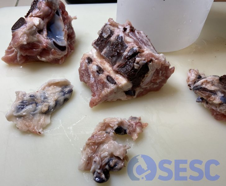
Samples were submitted from a 6,5 months old, hybrid pig carcass, which showed a darkish coloration of the bone marrow and the lymph nodes. Unfortunately, the viscera couldn’t be evaluated.
The samples were observed under the Wood lamp (UV) and revealed bright orange autoflorescense. Additionally, the histopathologic study revealed abundant brown extracellular material both on the bone marrow and lymph node.
All the findings regarding the characteristics of this pigment are consistent with congenital porphyria. Congenital erythropoietic porphyria affects multiple species such as bovine, swine, cats and humans and consists of a genetic defect that affects the metabolism of hemoglobin and leads to accumulation of the pigment in the skin, bone, teeth... depending on the species it can also produce anemia and/or photosensitization. Pigs only suffer from pigmentation in teeth and bone and uroporphyrin presence in urine, and never develop anemia or photosensitization. It is known that this condition is inherited with a dominant autosomal pattern but the specific genetic defect it is yet not known. (AC)
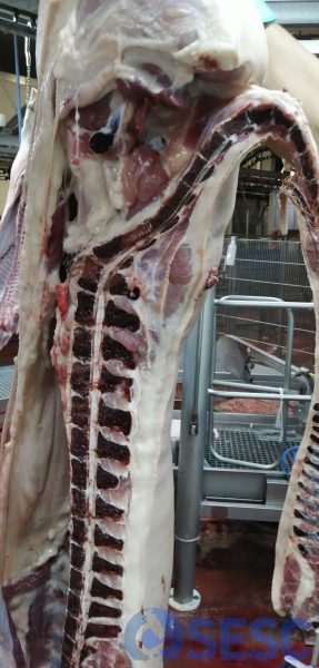
Half carcass of a pig which shows a darkened bone marrow.
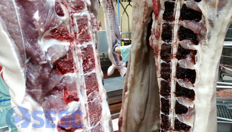
Comparison of the carcass with porphyria (right) with an unaffected carcass (left).
Section of the spine which shows the same appearance of the bone marrow. Also, a dark coloration of the cervical lymph nodes can be seen.
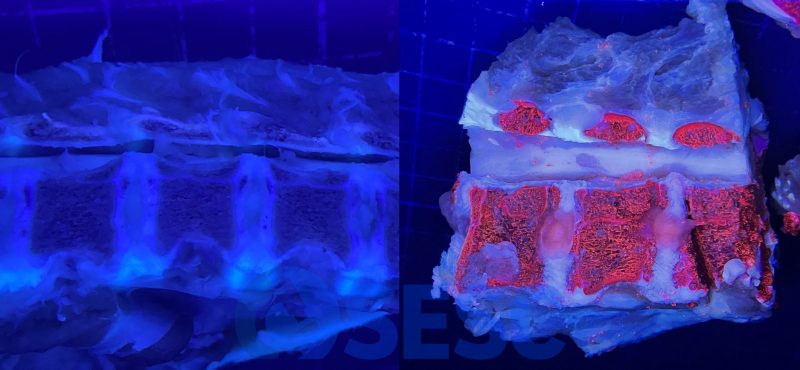
Vertebrae visualized under a Wood lamp, which reveals a bright orange coloration of the bone marrow. At the left, the vertebra of a normal pig, without autofluorescence.
