Lambs with sheeppox lesions
Two consecutive inquiries were submitted to SESC regarding two batches of lambs - coming from the same farm - with a series of skin and subcutaneous lesions. The affected animals are 4-5-month-old lambs, of the Manchega breed, and in the two batches, lesions were found in 2 and 7 animals respectively.
The lesions were nodular, prominent, and measured 1-2 cm in diameter, and involved both the skin and the subcutaneous tissue. When removed, they had a slightly red color and a firm consistency. The lymph nodes were slightly increased in volume. No lesions were reported on the rest of the viscera. Lymph node, skin and subcutaneous nodule samples were sent.
Histology was performed on all samples. In the skin, a marked vacuolization of the keratinocytes (balloon-shaped degeneration) was observed, with the presence of abundant intracytoplasmic eosinophilic inclusion bodies. Areas of ulceration (loss of epidermis) were also observed. The dermal and subcutaneous nodules presented a similar histological appearance, consisting of a dense macrophage and lymphoplasmacytic infiltrate, as well as a fibroblastic proliferation of intermediate cellularity (atypical mesenchymal proliferation). Abundant macrophages and fibroblasts in the subcutaneous tissue presented an intracytoplasmic inclusion body similar to that observed in the dermis. These cells receive the classic name of sheeppox cells.
These lesions are highly compatible with a case of sheeppox. It is a non-zoonotic disease, which affects sheep and has been considered eradicated in Spain since 1968. It is a disease included in the list of WAHO and of mandatory declaration in the EU (Regulation (EU ) 2020/2002) given its high transmissibility and mortality in naïve populations. Samples were immediately sent to the national reference laboratory for confirmation of the suspicion by PCR. Soon after, the infection was confirmed by a virus of the Capripoxviridae family (to be typified). In this genus there are two species of virus (sheep and goat pox, and contagious bovine nodular dermatitis). As many poxviruses generally do, capripoxviruses cause proliferative and necrotizing epidermal lesions. However, capripoxviruses are particular in terms of their presentation, as they induce very marked mesenchymal proliferations that characterize the lesional spectrum. The presence of pulmonary nodular lesions and other internal organs is also characteristic, although in our case they were not reported.
This case includes a suspicion and confirmation of a notifiable disease detected thanks to the slaughterhouse's Official Veterinary Services. The relevance of the passive surveillance carried out by the official inspection services should be highlighted, also with regard to diseases of importance in animal health.
After the epidemiological study, this case was linked to the outbreak recently declared in Granada. The competent authorities (DACC) have already launched active surveillance in farms that may be epidemiologically related without detecting symptoms of the disease nor viral circulation in Catalan farms. However, we want to take the opportunity to share the images of this case in order to keep the passive surveillance of this disease alert and reinforced since currently there are outbreaks declared in other spanish Autonomous Communities and Catalonia receives many animals from the rest of the state. That is why it is necessary to be alert to the potential appearance of new outbreaks in the event that this virus has circulated in the territory. (AC)
Link to the Spanish Ministry's website on Sheep and Goat Pox, with updates on the latest outbreaks.
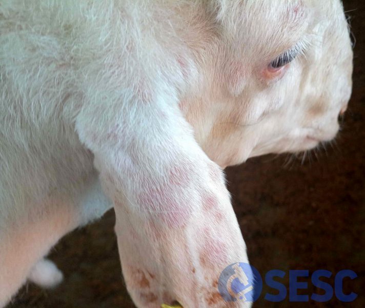
Nodular skin lesions on the face and ear of a lamb.
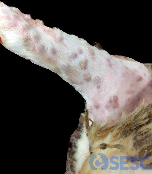
Slightly reddened nodular skin lesions on the hind limb of a lamb.
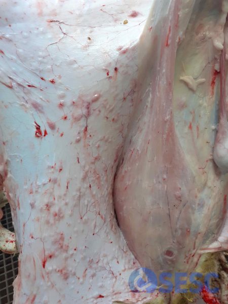
Skin lesions on the abdomen of a lamb, when removing the skin. It can be seen that the lesions bulge and affect the subcutaneous tissue.
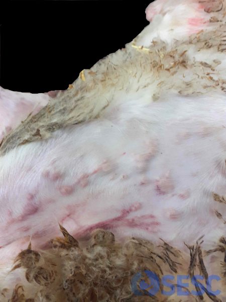
Nodular skin lesions in the ventral region of a lamb.
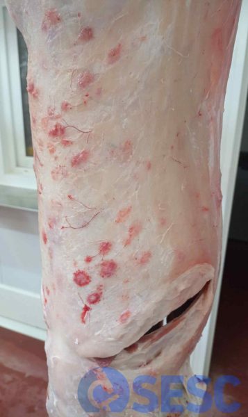
As nodular lesions affect the subcutaneous tissue, some of them may be retained in the carcass when the skin is removed. In this case, some have a very red halo, a product of the necrotizing nature of the lesion.
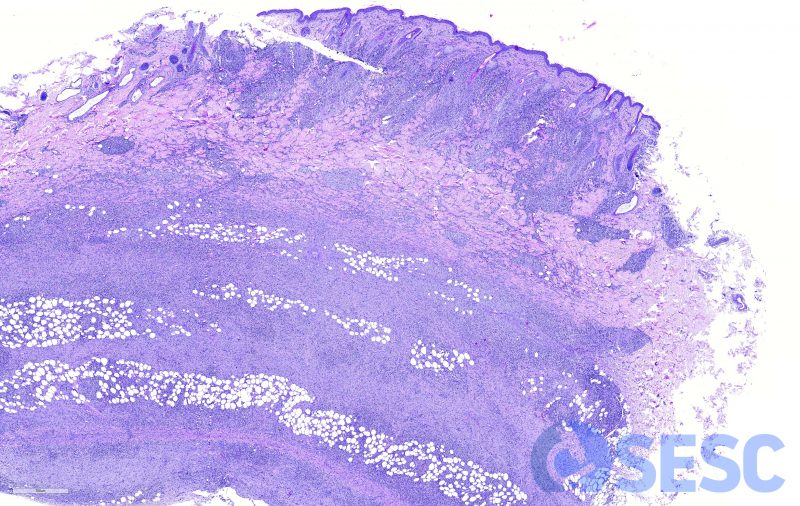
Histological section of a cutaneous nodule. Thickening of the skin due to moderate dermal and epidermal thickening can be seen, as well as marked cellular proliferation and inflammatory infiltrate in the subcutaneous tissue.
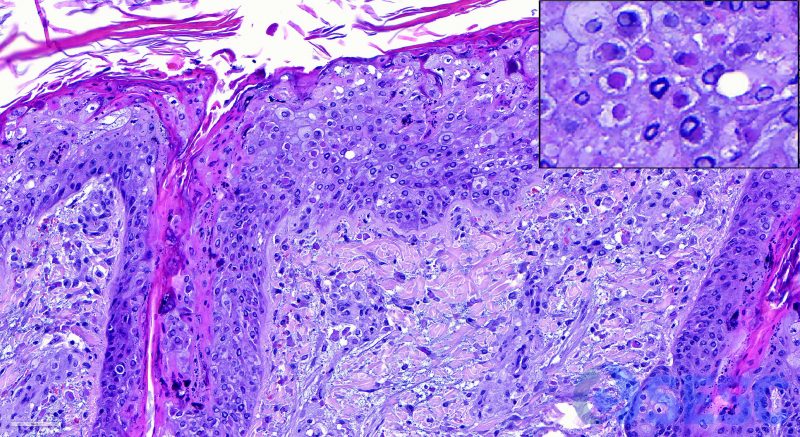
Detail of the epidermis. Vacuolization and swelling of the keratinocytes (ballooniform degeneration) can be observed. In many of them a single eosinophilic intracytoplasmic inclusion body is observed (top right insert).
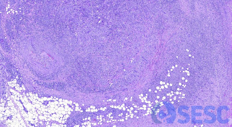
Nodular subcutaneous lesion. Histologically, a marked lymphohistiocytic inflammatory infiltrate is observed, as well as a mesenchymal proliferation that expands the subcutaneous tissue and infiltrates the adipose tissue.
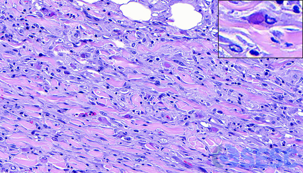
Subcutaneous mesenchymal proliferation. Among the normal collagen fibers, fibroblasts with loose nuclei and basophilic cytoplasm are seen, and frequently contain intracytoplasmic inclusion bodies (top right insert). Histiocytes with intracytoplasmic inclusion bodies are also observed. Both cells are called "sheeppox cells". Mesenchymal proliferation is often described as “atypical mesenchymal proliferation” given its histological characteristics. Both histological features (ovine pox cells and atypical mesenchymal proliferation) are characteristic features of capripoxviridae virus infections.
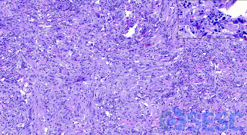
Detail of another area of atypical mesenchymal proliferation. In this case, an abundant lymphohistiocytic inflammatory infiltrate is observed. Macrophages often also have intracytoplasmic inclusion bodies. Karyolytic and karyorrhectic remains are also observed in some areas (necrosis, top right insert).
