Any given day in a rabbit slaughterhouse (2022)
Last June we organized a practical workshop on the identification and description of lesions for official meat inspectors. For this reason, we collected viscera and carcasses confiscated from several poultry and rabbit slaughterhouses, which were the subject of discussion during the session. In this post, the most frequent and/or interesting lesions are compiled, of which formalin samples were also collected for histological study. (AC)
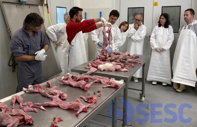
The pathologist and collaborator of the SESC, Carlos López-Figueroa, led the discussion of condemned rabbit carcasses and viscera.
CASE 1
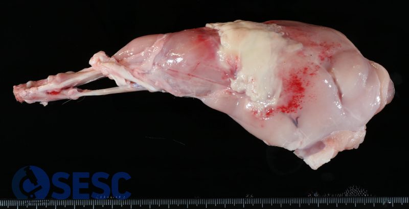
Rabbit limb. With a focally extense distribution, an accumulation of whitish material was observed in the fascia and muscle of the limb, which deepened in the section and was surrounded by reddened millimetric areas (petechiae).
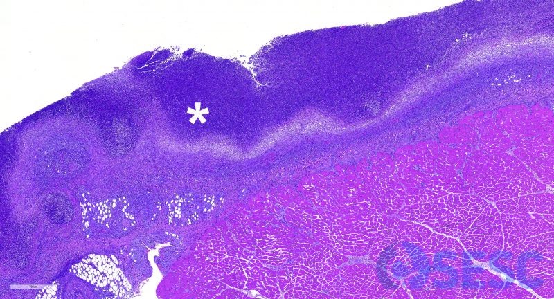
The histology corresponds to a heterophilic infiltration (*) of the muscle and fascia: myositis and subacute suppurative fasciitis.
These lesions in rabbits are usually associated with bacterial infections, usually by P. multocida, which gain access to the subcutaneous tissue through a wound. In these cases it is important to confirm that it is a localized infection and that there is no evidence of systemic dissemination (lesions or abscesses in other locations).
CASE 2
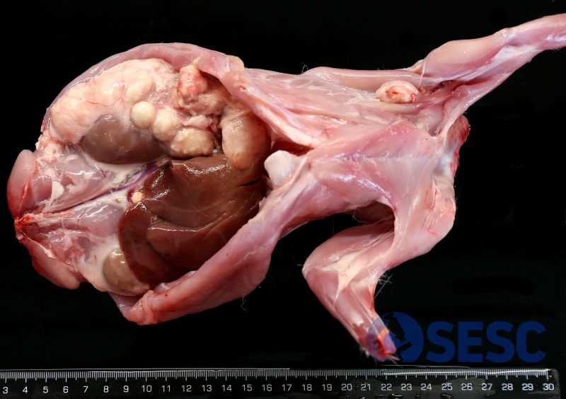
Rabbit carcass. In this case, multiple nodular lesions could be observed in the abdominal cavity, as well as in the armpit. Those in the abdominal cavity occupied a large part of the right retroperitoneal area, surrounding the right kidney, as well as adhered to the liver. All of them, in section, presented a pasty whitish content (pus) and a fibrous capsule 2-3 mm thick.
This is a common presentation of lesions produced with P. multocida infections, although other pathogens could also cause similar lesions (T. pyogenes, Staphylococcus spp...).
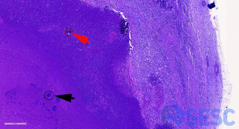
Histologically, the lesion corresponds to an abscess, that is, a collection of necrotic remains. On the left of the image, lytic necrosis (pus) is observed and in the center, viable and degenerated heterophils are recognized. Among this material, necrotic remains with dystrophic mineralization can be observed (red arrow), as well as occasional bacterial colonies (black arrow). This material is delimited - on the right of the image - by an inflammatory transition zone (with a predominance of foamy macrophages), surrounded by granulation tissue in different states of maturation, which finally becomes mature fibrous tissue, making up the capsule of the abscess.
CASE 3
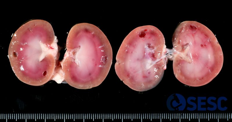
Rabbit kidneys. In this case, multifocal cystic formations were observed in both kidneys, which were found in both the cortex and medulla, and measured 1-4 mm in diameter.
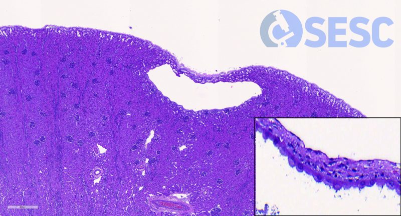
These lesions corresponded to renal cysts, filled with serous fluid, which derive from the dilation of the urinary space of the nephron (corpuscles and/or tubules). At the bottom right you can see that the cyst was lined by an attenuated cuboidal epithelium, evidence of the epithelium lining the tubule. Renal cysts can have multiple causes, they can be congenital or secondary to renal alterations - not observed in this case.
CASE 4
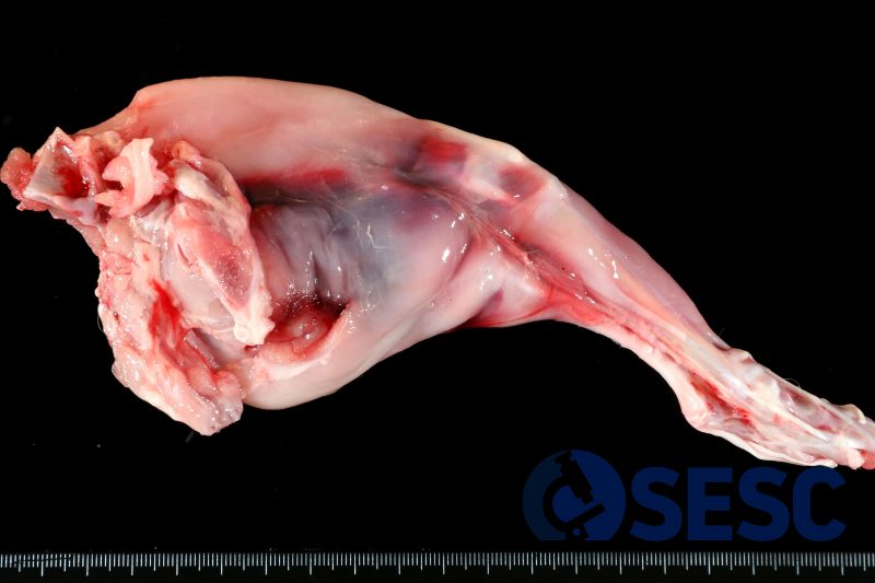
Rabbit hind limb. The thigh region is reddened and has a slightly gelatinous appearance in a focally extensive manner. At the opening, a hematoma (clotted blood) is observed between the muscle planes and the subcutaneous tissue.
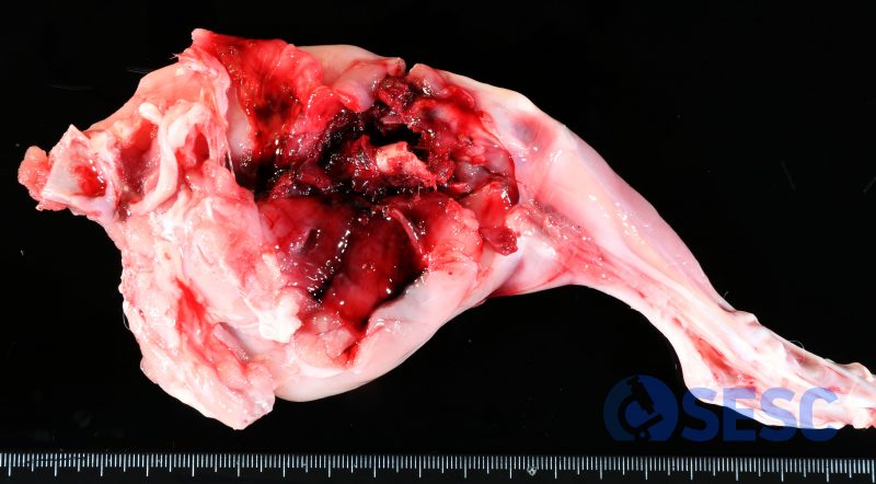
When dissecting the limb, a femur fracture was evident, which had caused the extensive bleeding.
These types of fractures usually result from handling the rabbit, for example, during the loading or unloading of animals and are an indicator of a lack of animal welfare in the slaughterhouse. Given its anatomy and behavior, holding the animal can cause sudden movements of the hind limbs, resulting in these fractures. Another common point of fracture in these situations is the spine, which usually results in posterior paraplegia.
CASE 5
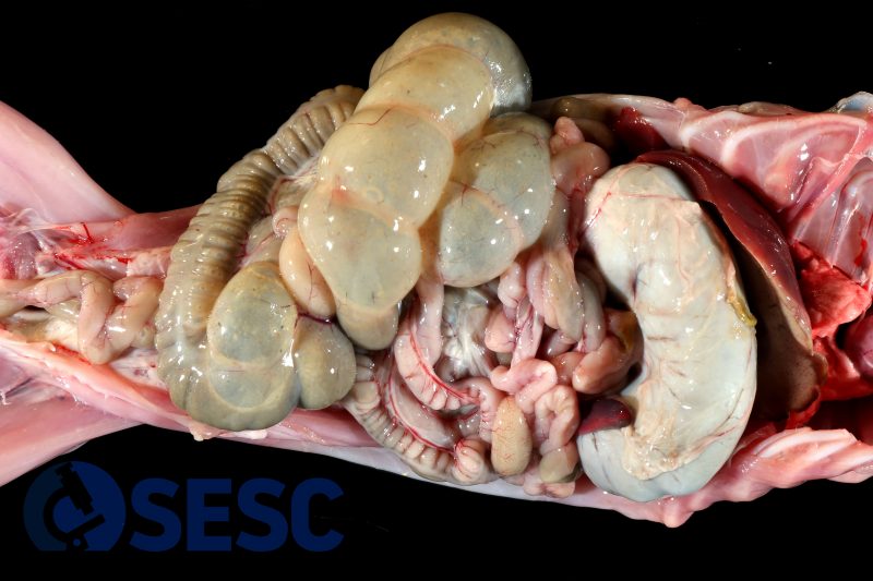
Rabbit large intestine. In this case, a marked distension of the large intestine (caecum and colon) could be observed with abundant gas inside.
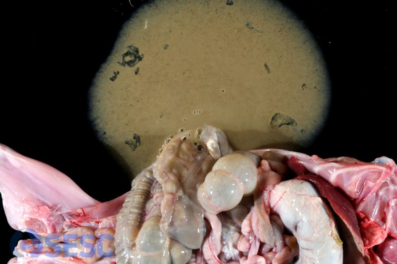
When you opened it, you could see how the contents were very fluidized (remember that at this point the content should be pasty). In addition, no stools were observed in the rectum and it had a pasty content, which is abnormal in rabbits. All these findings can be summarized with the morphological diagnosis of catarrhal enteritis, referring to the fluidification of contents.
In rabbits, reference is usually made to digestive syndrome in these situations, where the pathology is the result of a variable combination of agents and environmental factors. Among the agents we can find there, it should be noted that they can be bacterial (such as E. coli), viral (such as rotavirus, coronavirus), or parasitic (such as coccidia). In terms of environmental factors, all those that can alter intestinal motility are important, such as stress or diet (alterations in the fiber-protein ratio).
