Crateriform skin lesions on pig carcases
In this post we discussed the possible etiologies of skin lesions crateriformes observed in two pig carcasses.
- Case 1: 6 months old, male, hybrid breed pig carcass. Crateriform skin lesions with a generalized linear pattern.
- Case 2: 6 months old, hybrid breed, unspecified sex pig carcass. Necrosis of the tip of the ears and the tail. Crateriform lesions on the skin of the extremities.
In neither one of the cases the anomaly was detected in the antemortem examination , no alterations were detected in the other viscera of animals.
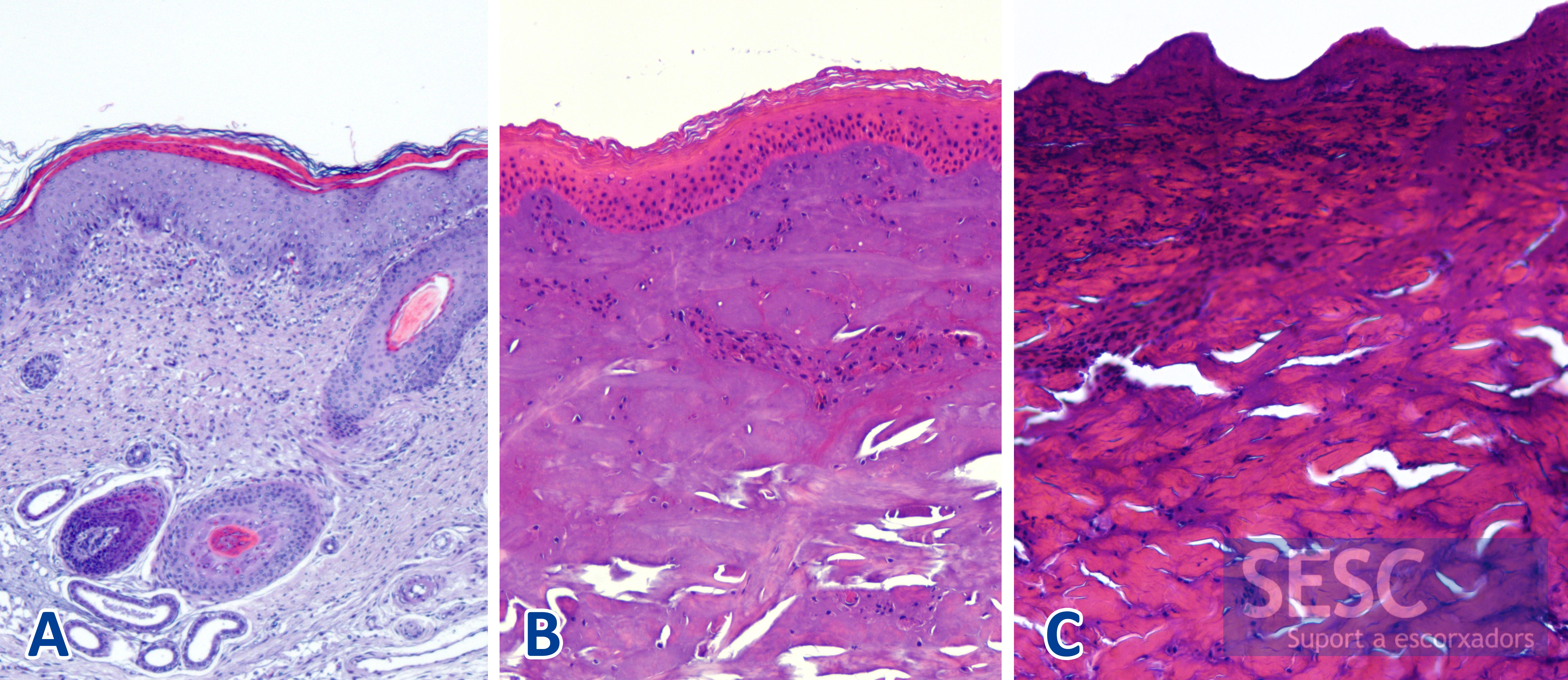
These lesions are compatible with sequelae of a crusty dermatitis among which on could include: pox, ptiriasis rosea, exudative epidermitis ... The possibility of actinic dermatitis sequelae (exposure to sunlight) was also considered. The problem in these cases is that the skin samples, once it has gone through the process of blanching, are artefactuated and do not allow a proper histopathologic diagnosis.
We invite you, if you have seen this type of lesion before, to leave your comment at the end of this entry.
Regarding the ear lesion it seems a case of the typical necrosis of the tip of the ears. The cause of which is always difficult to establish, especially if limited data is abvailable. It has been attributed to infections with Staphylococcus hyicus often over-infected with streptococci or spirochetes (sometimes all of it secondary to immunosuppression situations - for example at the time of the highest prevalence of PMWS, it was very frequently observed) or Mycoplasma suis. Moreover, it has been said that there are environmental-behavioral factors that predispose to ear-tip necrosis such as cannibalism, high concentration of ammonia, poor hygiene, mycotoxins, etc.
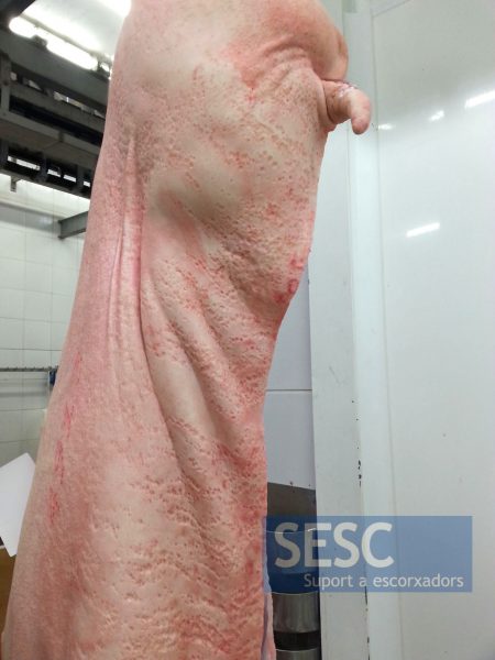
Case 1: Slight reddening of the skin and crateriformes lesions with a linear distribution throughout the carcass.
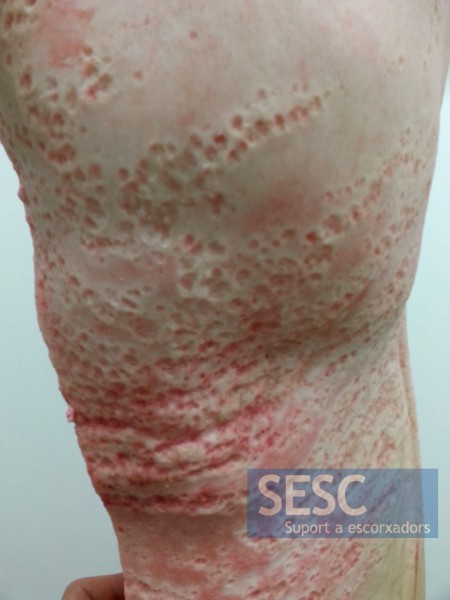
Case 1: Detail of the crateriform lesions.
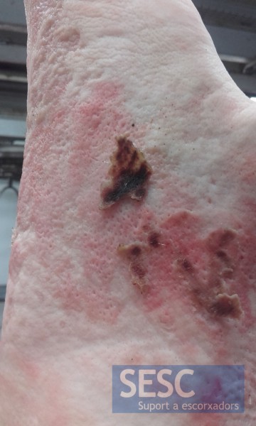
Case 2: crateriform lesions with crusty debris.
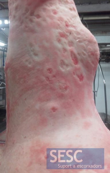
Case 2: crateriform lesions also affect the skin of the extremities.
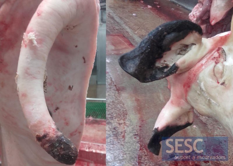
Case 2: This case also had necrosis of the tail and ears.

4 comment(s)
I have this sign’s at our piggery
Do you have images of the lesion before slaughter?
Comment posted in Linkedin’s Veterinary Pathology group:
By Kiril Dimitrov
No I have not seen such type of lesions, but few diseases may be related to this condition – severe Staphylococcal dermatitis (Staphylococcus hyicus or other spp.); Chronic erysipelas with skin necrosis; Chronic lesions due to dermatitis nephropathy syndrome; Swine pox infection; Mycotoxin related dermatitis or some kind of seborrheic dermatitis.
bacterial dermatitis and septicaemia.