Skin Neoplasia in a calf
In a cross-breed 10 months old calf, multiple nodular alopecic lesions were observed on the skin.
The histopathological study evidenced a neoplastic proliferation infiltrating the epidermis and dermis structures consisting of a population of round cells with abundant cytoplasm. In a few of them, brownish granules could be spotted. Infiltrating the neoplastic population, some eosinophil polymorphonuclear leukocytes could be observed.
Immunohistochemical markers for B (CD79) and T lymphocytes (CD3) were performed with a negative result. The toloudine blue stain did not allow to identify the presence of intracytoplasmic granules. Instead, C-kit marker gave a positive result in 100% of neoplastic cells.
The C-kit positive result and the presence of eosinophils infiltrating the mass support the diagnosis of a cutaneous mastocytoma. Negative results to lymphocytes markers help rule out a lymphoma. It is not possible however, to rule out that it is an amelanotic melanoma since these tumors can also react to the c-kit, but the presence of eosinophils tilts in favor of a mastocytoma.
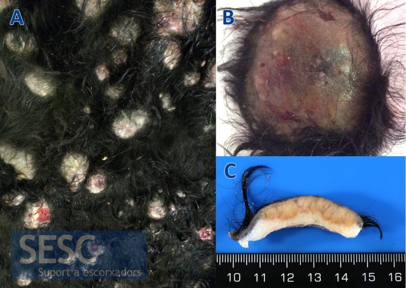
Gross appearance of the tumor. A: Multiple alopecic skin nodules. B: Detail of one of the nodules. C: Cross section of one of the nodules, the tumor invades the structures of the dermis.
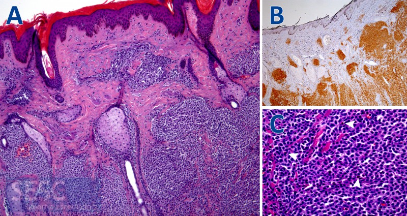
Histopathological study. A: H&E. Neoplastic growth infiltrating the structures of the dermis. B: Immunohistochemistry against the marker C-Kit, with a positive result in all neoplastic cells. C: H&E. Eosinophils polimorfonucelar leukocytes can be observed (arrowheads) infiltrating the neoplastic population.
