Diffuse cortical necrosis in pig kidneys
A case was submitted from a porcine slaughterhouse regarding one hybrid breed, 6-month-old pig carcass. At post-mortem examination, lesions affecting both kidneys specially caught the attention of the official meat inspectors, and for this reason they decided to submit one kidney to SESC for its anatomopathological study.
Macroscopically it presented lesions consisting of perirenal oedema and a diffuse whitish coloration of the renal cortex. Also, reddish lesions could be observed in the renal cortical surface (circular in shape), as well as a reddish rim in the renal corticomedullary junction. Additionally, the reddish rim extended multifocally from the corticomedullary junction radially into the cortical surface. This pattern is highly suggestive of a vascular process, as the renal vascularization follows the cortico-medullary junction (arcuate vessels) and the arterial ramifications extend from this region into the cortical surface (radiate vessels).
At histopathological evaluation of the lesions, the whole cortex exhibited acute necrosis with maintenance of the tissular architecture, i.e. it had suffered from diffuse acute coagulative necrosis. The coagulative necrosis is almost always product of situations of hypoxia/ischemia. In this case, it was accompanied by a generalized thrombosis in all the renal vessels and glomerular capillaries. Also, diffuse haemorrhage in the corticomedullary junction and delimiting the radiate vessels was observed (as noted macroscopically).
This lesion is known as renal cortical necrosis and consists in the necrosis due to ischemia of the entirety of the renal cortex. There are multiple possible causes, but it is always associated to a massive activation of the coagulation cascade (Schwartzman-like reaction). In all the species it is often associated to endotoxemia (presence of abundant bacterial toxins in blood) or shock. In swine it has been described associated to gastric ulcers, however it seems like the marked prevalence of gastric ulceration is swine isn’t accompanied by similar prevalence in the renal cortical necrosis (which is, in fact, rare). For this reason, although no other organs were studied hence, they probably did not present changes observable during post-mortem examination, it is considered that the most likely diagnosis is an endotoxemia. By means of precaution, it should be considered that the animal suffered from a systemic infectious process. (AC)
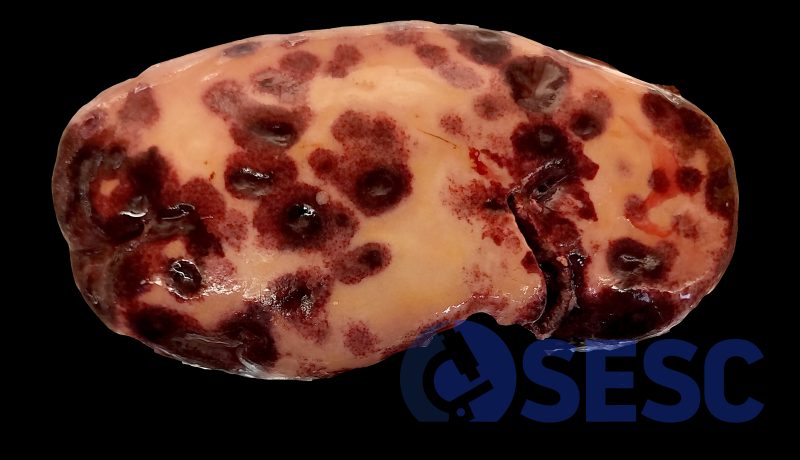
One of the affected kidney, with a very marked pale coloration of the cortex, as well as multifocal circular areas in the renal cortical surface.
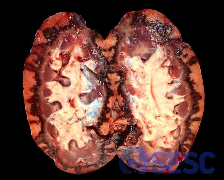
When sectioned, this pale coloration can be seen affecting the totality of the renal cortex, with the exception of the corticomedullary junction which shows a haemorrhagic appearance, with multifocal red streaks that extend into the renal surface (following the pattern of radial vascularization).
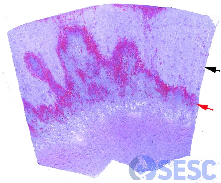
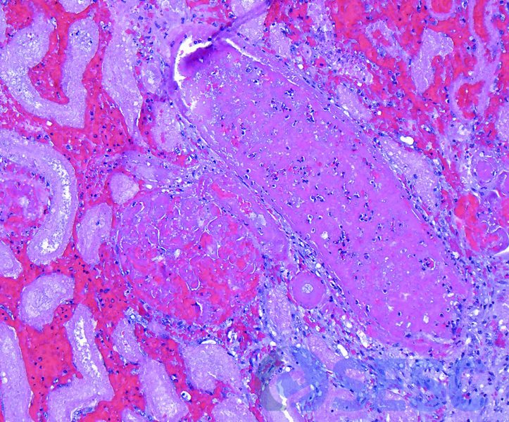
Histological image at low magnifications. The diffuse cortical necrosis can be observed (black arrow), as well as the haemorrhagic delimitation in the corticomedullary junction, with radial projections into the renal cortical surface (red arrow).
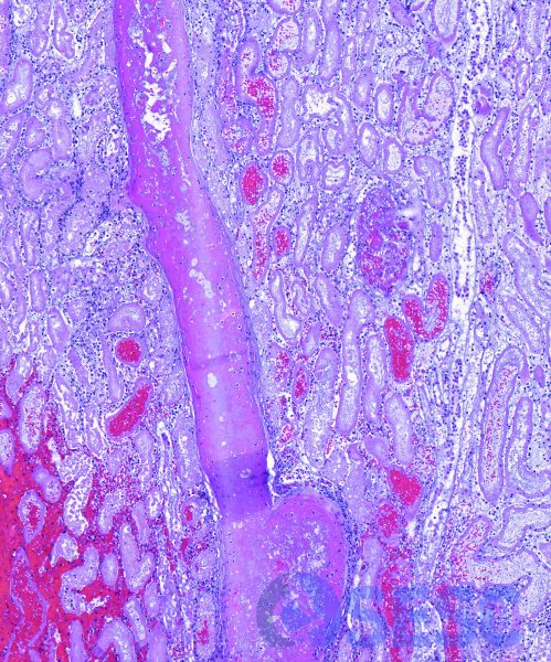
Abundant thrombosed vessels can be observed, especially arcuate and radiate vessels (following the haemorrhagic area).
Histologic detail of a thrombosed vessel, as well as a glomerulus that presents diffuse thrombosis of glomerular capillaries.

Occasionally some tubules in the medulla present fibrin within their lumen, indicating the exudation of fibrin from the glomeruli and its flow into distal parts of the nephron.
