22/11/2010
|
Abdominal cavity (porcine)
2
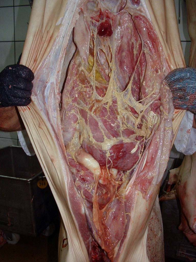
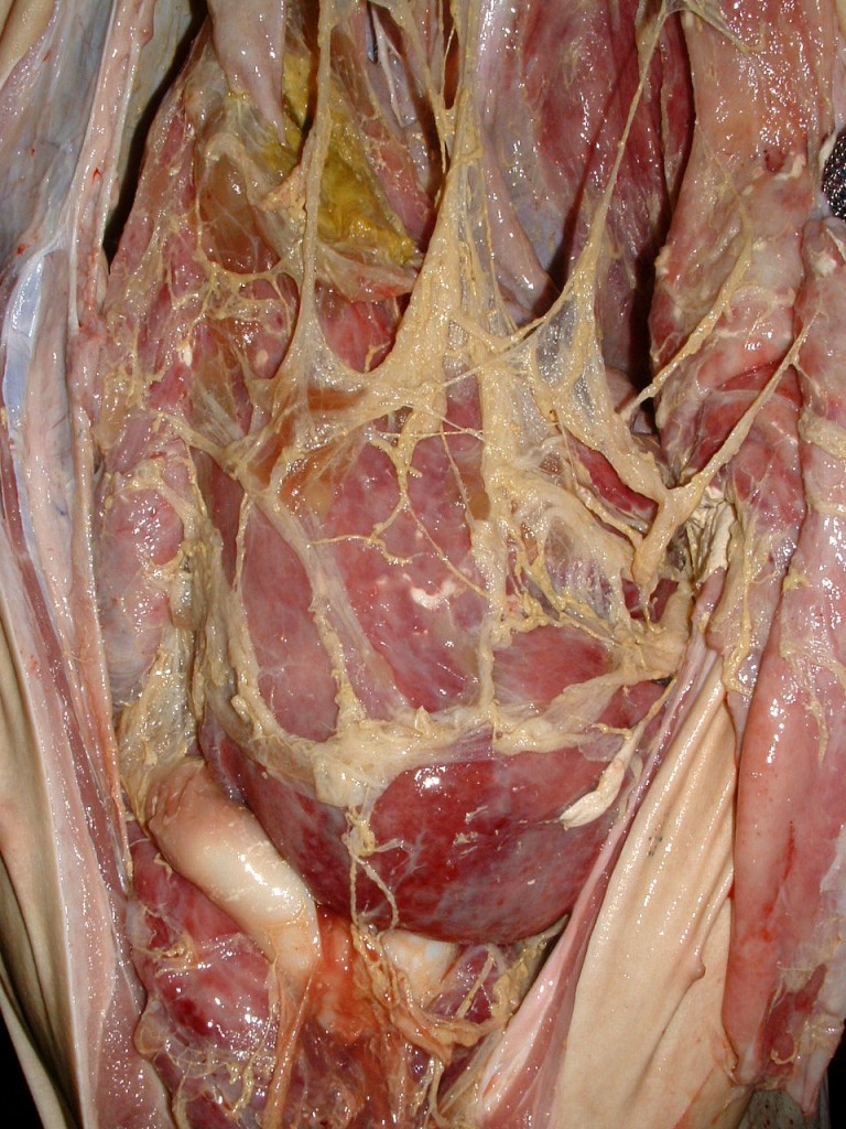
Fibrinous peritonitis in a pig carcass
In pigs, fibrinous peritonitis is often associated with systemic bacterial infections such as Haemophilus parasuis, Streptococcus suis or Mycoplasma hyorrhinis, and more rarely, Escherichia coli. Another possible cause of fibrinous peritonitis is perforation of the gastric wall.
A chronicity of this lesion could lead to a fibrous peritonitis where fibrin deposits are organized and are replaced by a proliferation of connective tissue creating adhesions between the viscera.


2 comment(s)
Should a carcass like this be condemned since this is again a chronic lesion.
When fibrin is still present, as shown in these pictures, it is indicative of an acute process. When chronic, this exsudate is arranged into fibrous strands (fibrous peritonitis) at this chronic stage one could consider the rest of the carcass fit for consumption since the causal agent is likely gone. But careful inspection should be conducted, including lymph nodes to ruleout an ongoing infection. Also, if the thoracic cavity is also involved (polyserositis) then condemnation due to generalized disease should be considered.