Follicular atrophy in a bovine carcass
An inquiry was submitted regarding a one-year-old Holstein calf. On ante-mortem examination, generalized redness of the entire skin with hair loss was observed. However, on post-mortem examination, no macroscopic alterations were seen in the viscera. The official veterinary service sent skin samples to the SESC for diagnostic purposes.
The skin samples were processed for histopathological study, through which multiple alterations were observed, mostly centered on the hair follicles. In these, diffuse atrophy is observed, with absence of hair and presence of disorganized keratin. Additionally, dilation of adnexal glands, dermal fibrosis, and perivascular lymphoplasmacytic inflammation are noted, findings interpreted as secondary to follicular atrophy. A diagnosis of alopecia due to congenital follicular dysplasia and secondary nonspecific dermatitis is made. It is also possible that the lack of hair made the animal more susceptible to solar radiation, contributing to dermal damage and inflammation. (AC)

Calf at the pre-mortem examination. The skin showed a generalized reddened coloration (erythema).
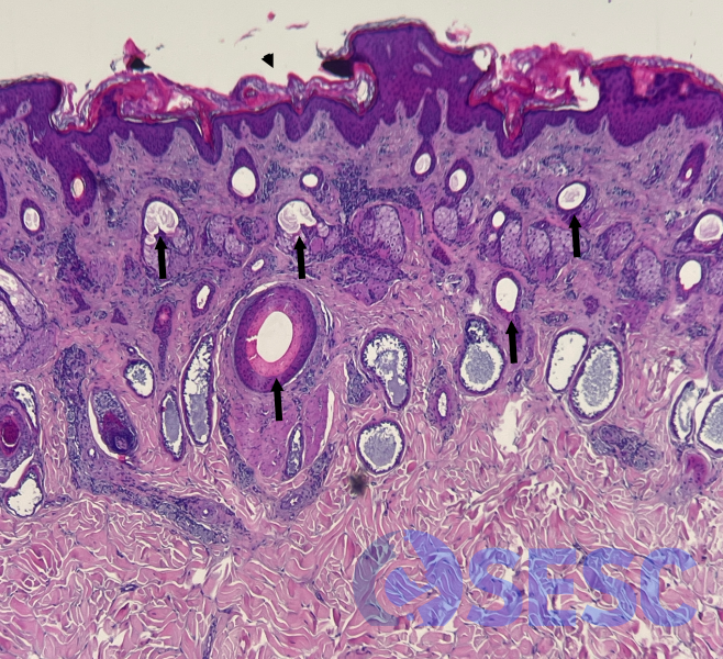
Histological section of the calf’s skin. Most of the follicles (both primary and secondary) showed no hair in their lumen, and most were in telogen phase (arrow). The epidermis showed marked hyperkeratosis
