Lesions in the mammary glands of sows
We have lately received a few enquiries related to changes in the mammary glands of female reproductive pig carcases. It is an organ which, in the case of recently weaned or lactating sows, can be allocated to human consumption.

Mammas of lactating sows stored for commercialization.
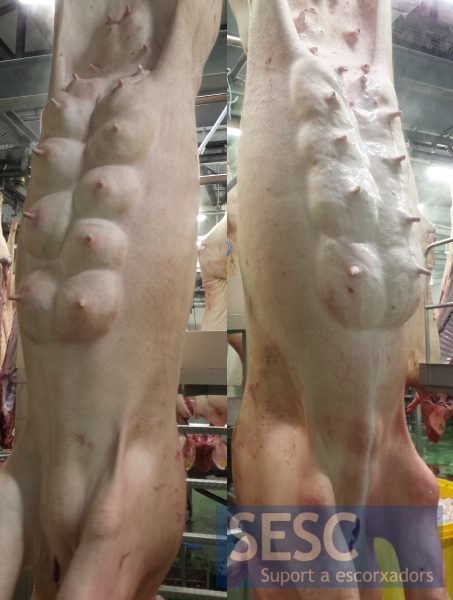
Sows usually have about seven pairs of mammas (3 to 10) in the thoracic, abdominal and inguinal regions. The thoracic mammas drain to the esternal lymph nodes while the inguinal and abdominal to the superficial inguinal lymph node. Each mamma consists of two separate units of glandular tissue (despite being independent glandular tissues they can be intertwined in one mamma) that drain into a single nipple via two separate conduits. There is no relationship between the different mammas. The arteries and veins that supply the mammary glands have anastomoses among them.
Different physiological changes can be observed in such carcases. It is normal to find mammas at different stages of hyperplasia or atrophy (dry mammas) in one animal, for example, mammas that have not been used by the piglets become atrophic. These are animals that are producing milk and when weaned, the excess milk has to be reabsorbed and can cause an increase in size and edematous appearance of the draining lymph nodes.
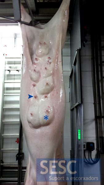
Reproductive sow carcass with mammas in different physiological states: glandular tissue atrophy (dry mammas) (arrow) and mammas still producing milk (asterisk).
As for the most common diseases that affect the mamary tissue of sows: acute mastitis in sows is usually caused by E. coli, S. aureus, Klebsiella spp. and by Streptococcus spp. to a lesser extent. Often more than one gland is involved and it is accompanied by systemic reactions (fever) and usually appear shortly after birth, associated with postpartum dysgalactia syndrome (formerly known as the mastitis-metritis-agalactia syndrome, MMA). Chronic granulomatous mastitis (formerly known as botryomycosis) can also bee observed, often caused by S. aureus, which can involve one or more mammas. Despite these microorganisms have been isolated from sows with mastitis they are also found in healthy sows so the etiology of mastitis is multifactorial (hygiene, hydration, immune status of the sow, etc ....).
A uniglandular mastitis (even of more than one gland) does not imply that other glands must have lesions because the glands are truly independent, especially in the case of bacterial mastitis where the entrance is through the nipple. It is understood that the rest of the carcass should not be affected either.
Instead, with pathogens arriving systemically such as Mycoplasma spp., Mycobacterium spp. or systemic fungal infections, for example, one would expect mutliglandular involvement. In these cases lesions should also be observed in the rest of the carcass/viscera as well as during the ante-mortem inspection (apathy, fever, etc.).
Other lesions often seen in sow's mammas are skin abscesses, associated with traumatic wounds or abrasions that piglets might cause during lactation. If the lymph nodes appear reddish it is an indication that, most likely, blood has been reabsorbed from their drainage area. In these cases a detailed inspection of the area is advised, including incision of the sternal and superficial inguinal lymph nodes to rule out the presence of additional lesions. We must also rule out the presence of lesions in other parts of the carcass that drain in these lymph nodes, such as an abscess in the posterior limb which drains into the superficial inguinal lymph node.
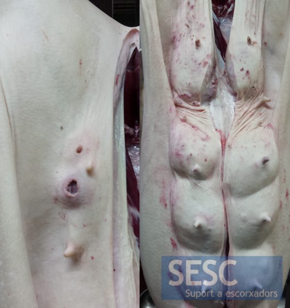
Examples of mammas with traumatic and/or abscessing skin lesions.
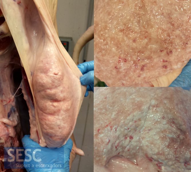
In one sow a mamma that had a hardened consistency was observed. When cut the mammary tissue was slightly reddened with small hemorrhagic foci. Histologically periacinar fibrosis and presence of neutrophils were observed in the interstitium and, to a lesser extent, inside the alveoli: mild chronic purulent mastitis. Although the lesion indicated a bacterial etiology, microbiological culture gave a negative result.
Two more carcasses with hardened mamas were sampled. Histologically both had mild periacinar fibrosis but no active mastitis lesions. However, microbiological culture of one of the mammas was positive since abundant beta-haemolytic colonies were isolated identified as Streptococcus Group L (which includes Streptococcus dysgalactiae, a bacterium that can be a commensal or cause mastitis in cattle and pigs).
