

Sampling of lymph nodes in bovine carcasses for the diagnosis of tuberculosis
Within the framework of the bovine tuberculosis (TB) eradication program, the Spanish Ministry of Agriculture, Fisheries and Food requires the sampling for tuberculosis diagnosis of in all those animals that have tested positive for skin tests (intradermotuberculinization) and are, thus, slaughtered. The objective is to confirm the disease and isolate the mycobacterium in order to carry out molecular epidemiology studies that will help us understand the origin of each outbreak: residual infection, contact with domestic or wild reservoirs, etc. And thus, be able to make the best decisions in each case.
The control of tuberculosis is very complex and depends on many factors for the outcome of the program's efforts to be successful. In this post we want to review, in the context of the veterinary inspection of carcasses, the sampling of target tissues in order to ensure that this will result in maximum diagnostic sensitivity. On some occasions we have detected some incidences and we will comment on the most common ones and the anatomical location of the different samples.
The Ministry published a sampling manual that you can consult here (in Spanish).
The sample of choice for TB diagnosis are tissues with granulomatous lesions, therefore when it is necessary to sample animals sacrificed due to unfavourable results in the in vivo tests, an additional effort must be made to locate these lesions. TB can be limited to small millimetre lesions that are impossible to detect macroscopically or, at the other extreme, we can find very large lesions or generalized patterns such as pearly tuberculosis. When we do not observe visible lesions, we must carefully palpate the lungs and inspect the animal's lymph nodes (NL) as closely as possible. In case of lesions, the sample that must be submitted is the lesion with a little adjacent healthy tissue. In an infected animal, mycobacteria will be found wherever there are lesions, so this sample is the one that has the greatest guarantee of diagnostic success.
In the case of animals without visible lesions compatible with TB, the manual indicates that samples must be taken from the following lymph centers:
- Head: retropharyngeal NL and submandibular NL
- Thorax: caudal mediastinal NL and left tracheobronchial NL
- Thoracic limb: pre-scapular NL
- Abdominal cavity: hepatic NL and mesenteric NL
- Mammary gland: supra-mammary NL
In bold we have marked the most relevant in the pathogenesis of tuberculosis in cattle.
Sampling of the retropharyngeal NL (NL RF).
Mistakes to avoid:
- Sampling of only one of the two lymph nodes (reduces the possibility of detecting lesions).
- Sampling, by mistake, of the palatine tonsil.
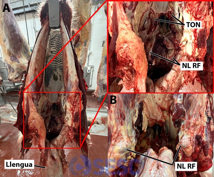
A: bovine head separated from the carcass, hung by the jaw and presented for inspection of the masseter muscles and LN RFs. The NL RFs are located dorsally in the pharynx. In this presentation the tongue and pharynx have been removed ventro-caudally after sectioning the soft palate, leaving both the NL RFs and the palatine tonsils (TON) exposed. B: Sometimes, as in the image, if the cut in the palate is deep enough, the NL RFs can be more external, on the sides of the base of the tongue.
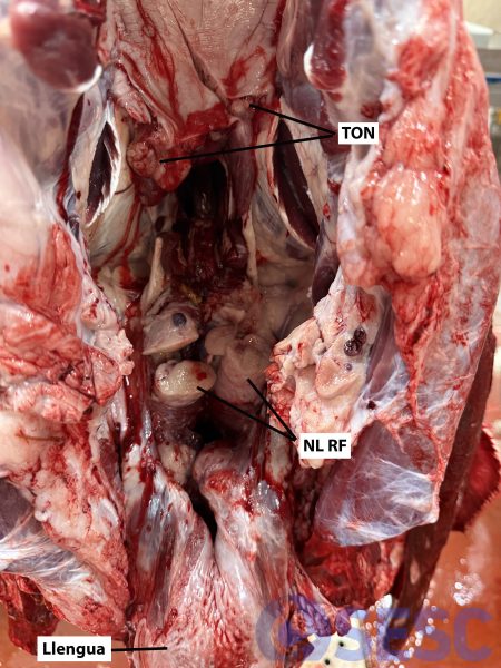
Detail of another example of a bovine head presented for inspection hanging from the jaw: the tongue and pharynx have been removed ventro-caudally exposing the RF NLs (in the image they are sectioned) and the palatine tonsils (TON), also sectioned, in the upper part, on the soft palate.
Sampling of the thoracic NLs: left tracheobronchial (NL TB) and caudal mediastinal (NL MD).
Mistakes to avoid:
- Sampling by mistake of the cranial mediastinal NL (less representative).
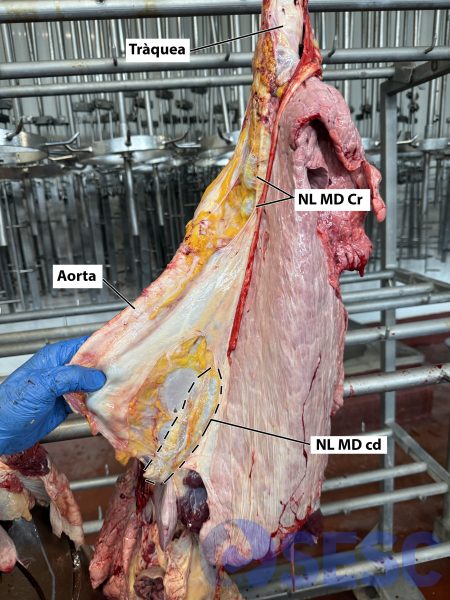
Bovine lungs hanging from the trachea: if we pull the aorta, which is located in the mediastinum -space between the two lungs-, we can find the caudal NL MD (NL MD cd), it is located in the serous membrane that separates the two hemithoraxes in the most caudal part, close to the diaphragm. The NL MD cd is elongated (it can measure about 10-15cm), it is important not to confuse it with other smaller NLs that are in the mediastinum and that are found more cranially (NL MD Cr) that are less representative of the lymphatic drainage of the lungs.

Pulmons de boví penjats per la tràquea: per localitzar el NL TB esquerre cal retirar cranialment l’aorta (i l’esòfag, si ha quedat enganxat) tal i com es mostra a la imatge i localitzar la bifurcació de la tràquea. El NL TB esquerre es troba entre la tràquea i l’aorta i el bronqui esquerre, en el punt on la tràquea es bifurca. Si l’animal té un bon estat de carns és normal que estigui rodejat de teixit adipós i costi de trobar.
Sampling from the abdominal NLs: mesenteric NLs (NL MS) and hepatic NLs (NL H).
Mistakes to avoid:
- Sampling by mistake of the adrenal gland (instead of the hepatic NL).
- Undersampling of the mesenteric NL (if we only sample a single small nodule it is unlikely that we will find lesions, the most elongated portion should be located and sampled).
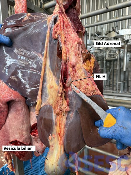
Bovine liver hanged for inspection: the visceral side is shown, the knife points to the NL H. Care must be taken not to confuse it with the adrenal gland (Gld Adrenal) which may remain attached to the liver when it is extracted from the abdominal cavity..

Bovine liver: detail of the NL H (sectioned in the image) where a light grayish cortex and a blackish medulla can be observed. In the upper part we observe the adrenal gland (Gld Adrenal), also sectioned which presents a light-colored medulla and a brownish cortex. In case of doubt as to whether a structure is a NL, it can always be sectioned and the characteristics of the parenchyma can be observed.

Bovine digestive system. To locate the NL MS, it is necessary to extend the mesentery, the serous membrane from which the intestines hang, and locate the chain of lymph nodes found in this membrane. We recommend sampling the longest one, since if we only sample a small nodule, the probability of detecting lesions is reduced. They are usually covered with adipose tissue, which makes their location difficult. Ru: rumen; Ab: abomasum; Om: omasum; Re: reticulum; Je: jejunum and Ce: cecum.

Another example of NL MS in cattle. When there are fewer fatty reserves, it is easier to identify them.
Sampling of pre-scapular NL (NL PE) and supra-mammary NL (NL SupMa).
Mistakes to avoid:
- Sampling of only one of the two lymph nodes (reduces the possibility of detecting lesions).

Bovine carcass: to locate the NL PE, the muscles in front of the shoulder (Espatlla) must be dissected with a knife, the NL is located below the trapezius muscle. Elbow: cloze.

Bovine truss separated from the carcass: the NL SupMa are located on the gland in the most cranial part, they are rounded and flattened.
Sampling for the diagnosis of paratuberculosis.
Paratuberculosis is caused by Mycobacterium avium paratuberculosis and sometimes this infection can interfere with tuberculosis diagnostic tests. In some cases, samples are requested to rule out this pathology that presents with thickening of the intestinal wall, lymphangiectasia and lymphadenomegaly of the mesenteric and ileocecal lymph nodes, generally, due to the presence of a diffuse granulomatous inflammatory infiltrate, although in severe cases it can form nodular granulomas similar to those of tuberculosis.
The sample of choice is the lymph node (NL IC) and ileocecal valve (VIC), although for diagnostic purposes sampling a fragment of about 10 cm of the distal ileum is sufficient. It is important that it is the distal ileum (close to the valve) since there is abundant gut-associated lymphoid tissue (GALT) there, which is where we will initially see the lesions.
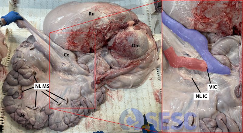
Bovine digestive system: to locate the ileocecal valve (VIC), it is first necessary to identify the cecum, grasp it and observe it from one side to the other until the point where the most distal part of the ileum (last section of the small intestine) is inserted into the cecum is visualized, this point is the ileocecal valve. In the enlarged photo on the right, we have marked the cecum in blue and the last fragment of ileum in red (this red fragment would be the target sample for diagnosis, ideally, before placing it in the container it is advisable to clean the fecal content with water). The ileocecal lymph node (NL IC) is very close to the valve, the fat around it must be dissected to locate it. In the image on the left, we can also see the sectioned mesenteric lymph node (LN MS). Ru: rumen, O: omasum; je: jejunum, Ce: cecum.
What do we do when the samples arrive at the laboratory?
The samples must be sent refrigerated to the laboratory as soon as possible to avoid the growth of contaminating microbes that may interfere with microbiological tests. If it takes more than 48 hours, it is advisable to freeze the samples to avoid autolysis, although freezing can reduce the number of viable mycobacteria and also hinders the sample quality for histopathological studies.

In the laboratory, a serial sectioning of all the lymph nodes is performed, 3mm thick, with the aim of detecting lesions that may have been overlooked during post-mortem inspection. If any are found, they are confirmed to be compatible with tuberculosis in the histopathological study and acid-resistant bacilli are sought with Ziehl Neelsen staining. A sample of the lesion is also taken to perform PCR for mycobacteria and microbiological culture. If there are no visible lesions, a fragment of an apparently healthy RF, TB and MD lymph node is taken for histology, PCR and culture.

Bovine pre-scapular lymph node: detection of an encapsulated lesion with a suppurative caseous appearance (arrow). In this case, it is classified macroscopically as “Inconclusive” since it is a lesion that is not very characteristic of tuberculosis, but could be compatible. The histological study showed that it was a pyogranulomatous lesion with the presence of bacterial colonies and tuberculosis was ruled out.
Finally, a reminder that this sampling procedure applies to animals that the agriculture department indicates to be sampled for the application of the bovine tuberculosis surveillance program, either because they have obtained unfavourable results in field tests or because they are follow-up cattle, but it is necessary to remember another very important surveillance component of the official slaughterhouse inspectors, which is the passive surveillance, that is: detecting and submitting for analysis the granulomatous lesions found in animals that have not been diagnosed while alive. To achieve the objective of analysis of 0.17 non-tuberculous granulomas for every thousand animals slaughtered set by the Ministry of Agriculture, in Catalonia, it would be necessary to analyse about 85 non-tuberculous granulomas each year. For this reason, it is important to palpate lungs and section lymph nodes to find potential incipient lesions.
☣️Biosafety warning: Tuberculosis is a zoonosis, that is, it can affect humans. Whenever samples are taken from animals that have tested positive for TB, the use of personal protective equipment (gloves, goggles and mask) is needed. In case of handling tuberculous lesions, where mycobacteria are found, the glove must be considered contaminated and disposed of appropriately (biohazardous waste).(EV)