Pig carcass with nodular lesions in the pleura and viscera
Although teh appearance was not of caseous necrosis the inspectors suspected of a tuberculosis case.
The histopathological study showed that it was a malignant round cell neoplasia: lymphosarcoma. In pigs, tuberculous lesions have a proliferative nature, rather than necrotic and, macroscopically, may resemble the aspect of a neoplasia. Therefore, it was necessary to rule out that it was an outbreak of tuberculosis. Tuberculous lesions in pigs can be caused by mycobacteria within the M.tuberculosis complex and M.avium.
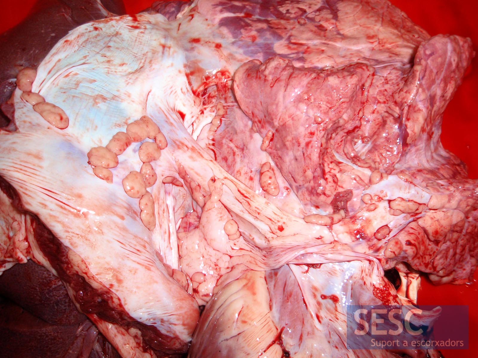
Whitish nodular lesions in the parietal pleura and lung.
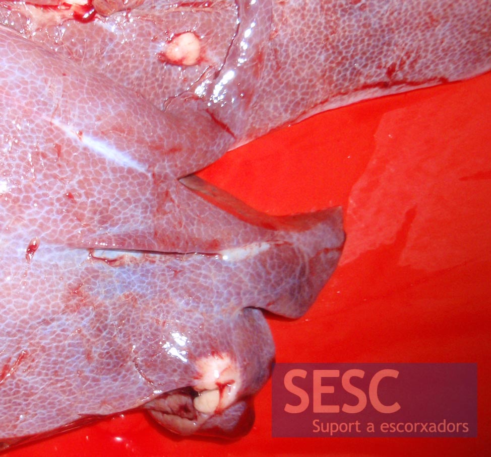
Whitish nodular lesions in the liver.

Image of Histopathology of the liver showing a proliferation of round cells (upper right, bluish). At higher magnification (inset) one can observe neoplastic cells.

2 comment(s)
Thanks for your comment Martín, we had a few cases of M.avium in pigs a couple of years ago:
https://sesc.cat/lesions-granulomatoses-disseminades-en-canals-de-porci/?lang=en
Then the suspected origin of the infection was the feed fed to the animals, which was the dame in the different farms that has cases.
comment from the LinkedIn group: Swine Health, Nutrition and Production Professionals
Martín Liebstein
Clinical Research Veterinarian at RTI, LLC
There has been a recent conversation within some swine veterinarians in the US, about pigs being condemned at slaughter with similar findings to this case you have posted. Apparently, the inspector at the plant determined that the animals had lymphosarcoma. Further testing done by the company’s vet has shown that the swollen lymph nodes correlate to PCV2 diagnosis. Other vets suggested that M. avium could be playing a role, introduced by birds getting in contact with the pigs in the barn. Definitely, different diseases to be considered and a good reminder to keep our eyes and mind open.