Any given day in a poultry slaughterhouse (2022)
Last June we organized a workshop for the identification and description of poultry lesions for official meat inspectors. For this reason, condemned viscera and carcasses were collected from poultry slaughterhouses, which were discussed during this session. In this post we discuss the more frequent and/or interesting lesions, from which samples were fixed in formalin for histopathologic assessment. (AC)
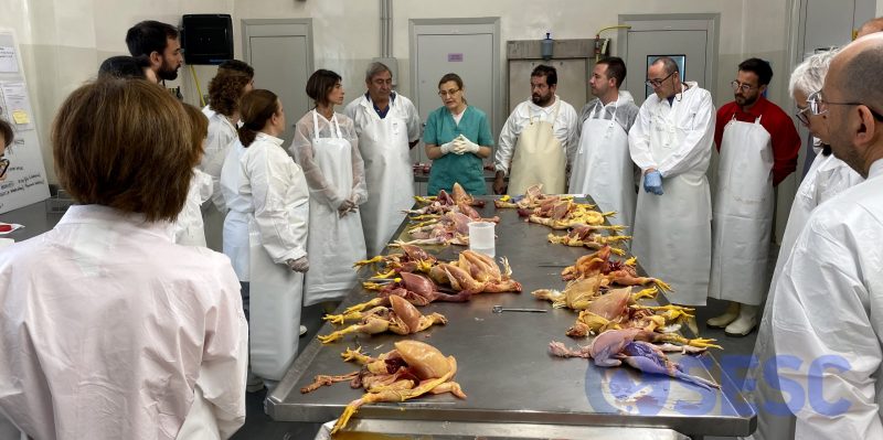
Natàlia Majó, Miquel Nofrarias and Carlos López (SESC colaborator pathologists) led the discussion session.
CASE 1
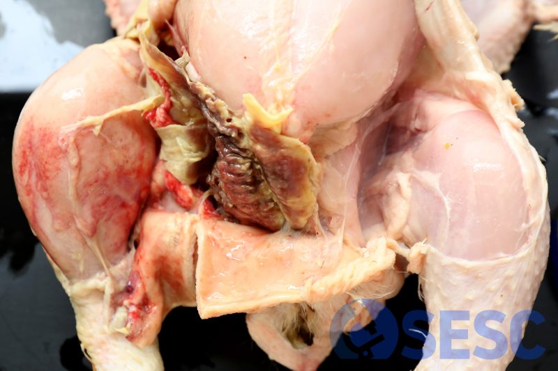
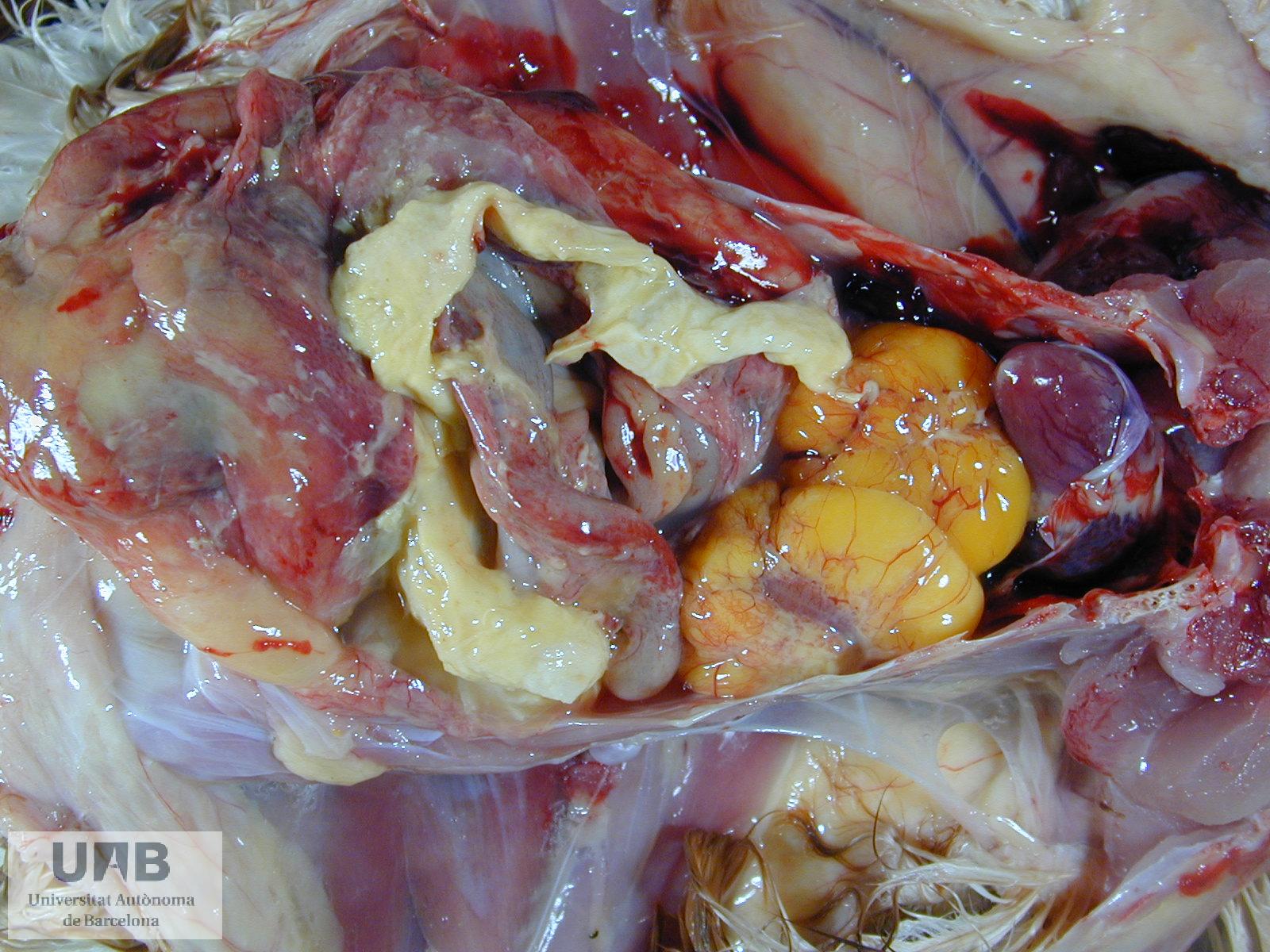
Broiler carcass. Externally showed an area of tumefaction and induration with darker coloration in the region of the thigh and abdomen (not shown in the images). When the skin was removed, yellow-green material could be seen occupying the subcutaneous tissue (between the skin and the muscle), as well as petechiae in the adjacent regions.
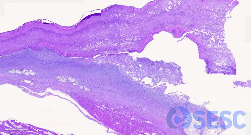
At histology, this material corresponded to necrotic debris, degenerated heterophils and fibrin, which separated the muscular tissue from the skin. These lesions were identified as fibrinoheterophilic cellulitis and are generally caused by skin lesions (scratches) and secondary contamination with bacteria from the environmentMost of the times E.coli is isolated, but can also be caused by Aeromonas spp. or P. aeruginosa among others.
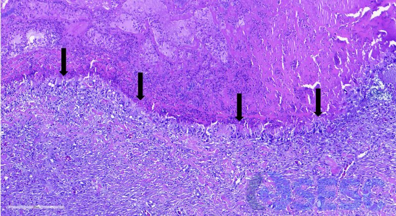
At histology, the necrotic debris were well demarcated by a thin rim of epithelioid macrophages, occasionally multinucleated (arrows).
This reaction, in mammals, would be defined as granulomatous. Nevertheless, heterophils in birds (equivalent to mammal neutrophils) lack the enzyme myeloperoxidase, which results in heterophilic and fibrinous exudates frequently undergoing caseification instead of liquefaction (in other words, birds don’t have pus). These necrotic debris are surrounded by macrophages and multinucleated giant cells which intend to slowly phagocytize and remove this caseificated material. For this reason, general pathology nomenclature is adapted in birds to try to adjust to the differences between exudates. Some examples are:
| Mammal term | Poultry term |
| Suppurative or purulent | Heterophilic o granulocytic |
| Pyogranulomatous | Heterophilic granuloma |
| Granulomatous | Histiocytic granuloma |
| Fibrinopurulent | Fibrinoheterophilic |
CASE 2
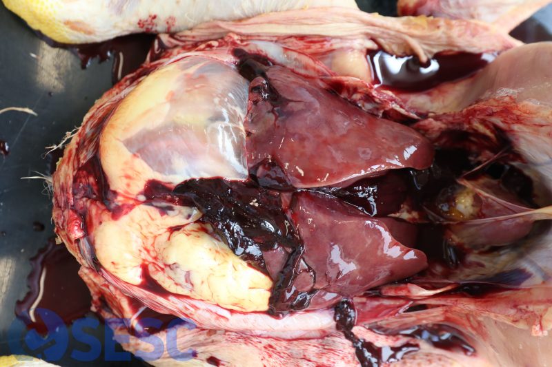
Broiler carcass. When opening the coelomic cavity, free blood could be seen within the cavity as well as clotted blod, specially around the liver.
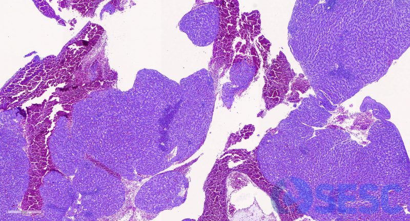
Histology confirmed focally extensive disruption of hepatic capsule and associated hemorrhages, as well as mild hepatic lipidosis.
These findings are frequently refered to as hemorrhaghic fatty liver syndrome, which refers to the increase in friability of the liver as a consequence of the fatty change. In this situation, trauma which would otherwise be harmless result in this intracelomic hemorrhage which leads to sudden death. When observed at slaughterhouse it is normally associated to transport related traumatisms.
CASE 3
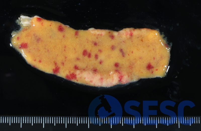
Broiler intestine. In this case the intestinal mucosa showed multifocal 1-4mm round reddish areas.
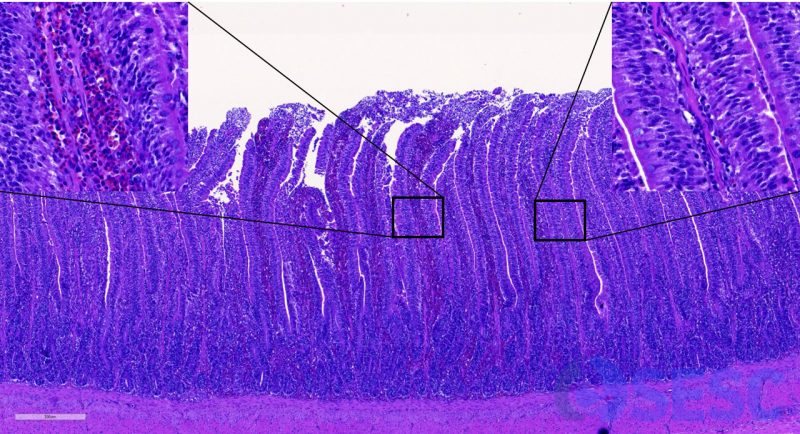
Histologically corresponded with extramedullary hematopoyesis in the intestinal villi. This finding is relatively common in young birds and doesn’t have any pathological implication.
CASE 4
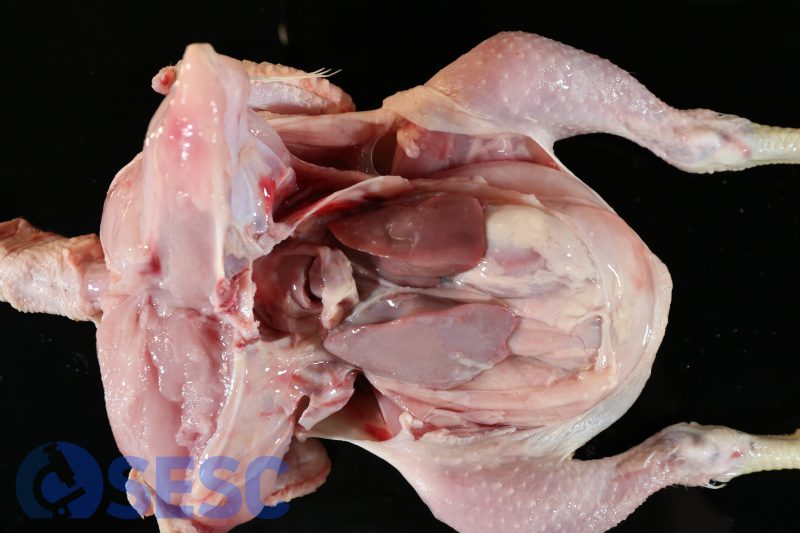
Broiler carcass. In this case, a marked celomic distension could be seen due to the presence of abundant intracoelomic liquid. Additionally, the liver appeared pale and slightly indurated.
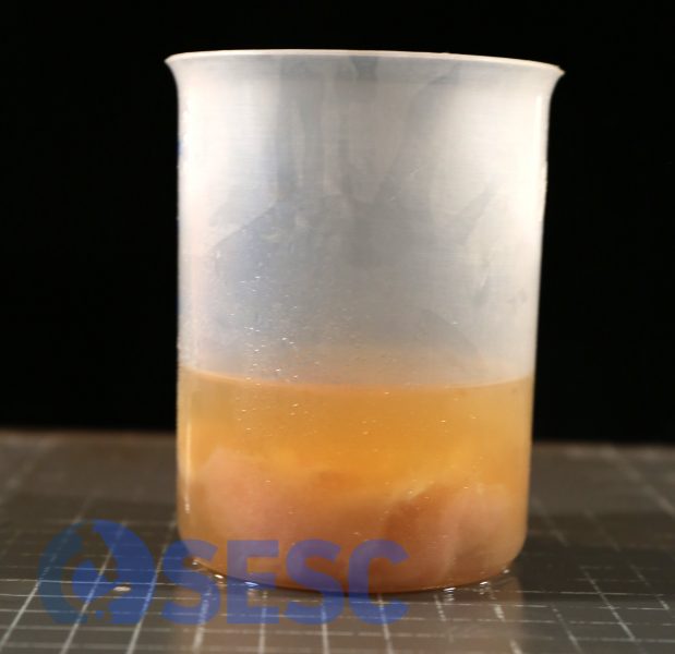
Intracoelomic liquid collected from the carcass, which was transparent with yellowish coloration and with abundant fibrin clots.
The right ventricle was dilated (in a healthy animal the right ventricular lumen should be almost virtual).
These are typical findings of broiler ascitic syndrome case. The pathogeny of this condition consists in an increase of the cardiocirculatory needs due to the rapid growth of broilers. As the lungs are located in a narrow space between the ribs and have limited expansion capacity, the excessive cardiac effort results in pulmonary hypertension, which retrogradely affects the right heart generating a congestive right heart insufficiency, which results in ascites and hepatic passive congestion.
CASE 5
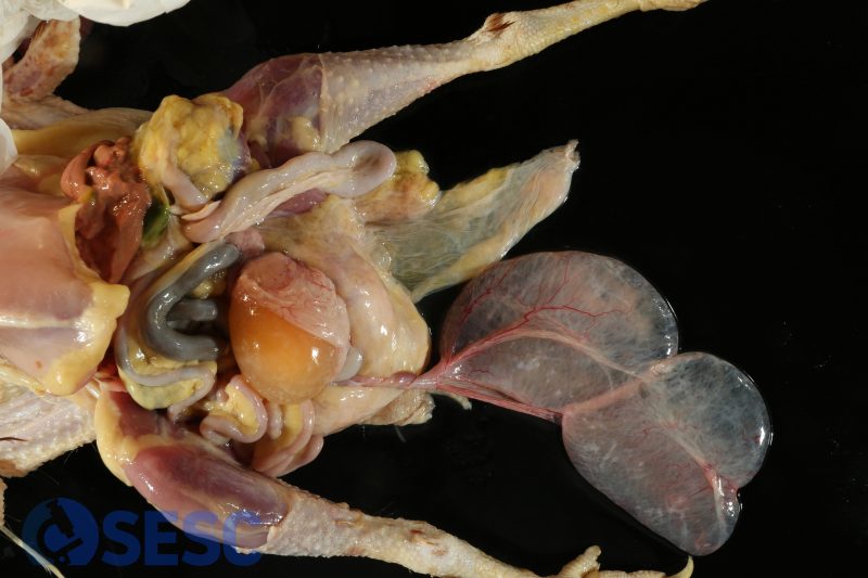
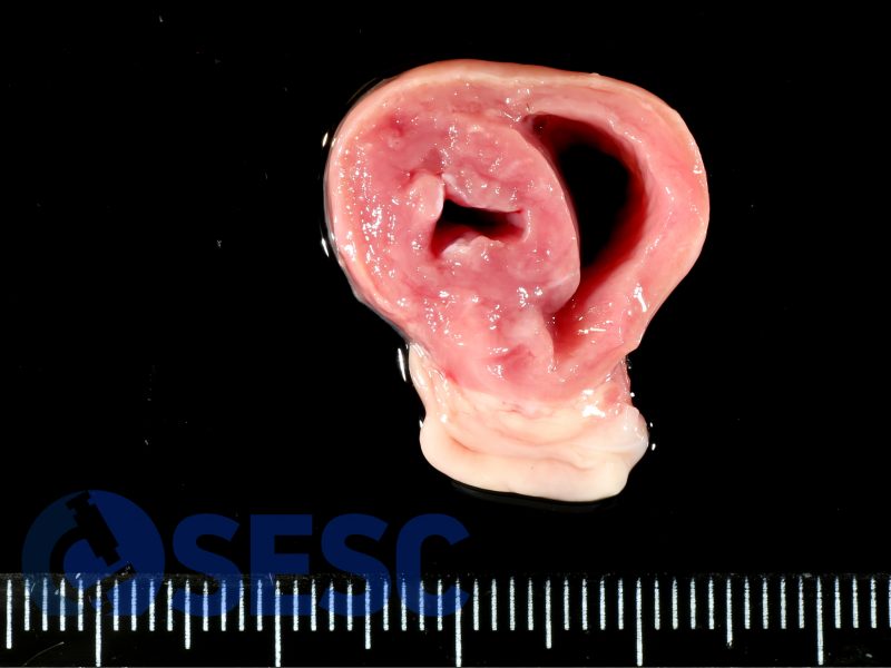
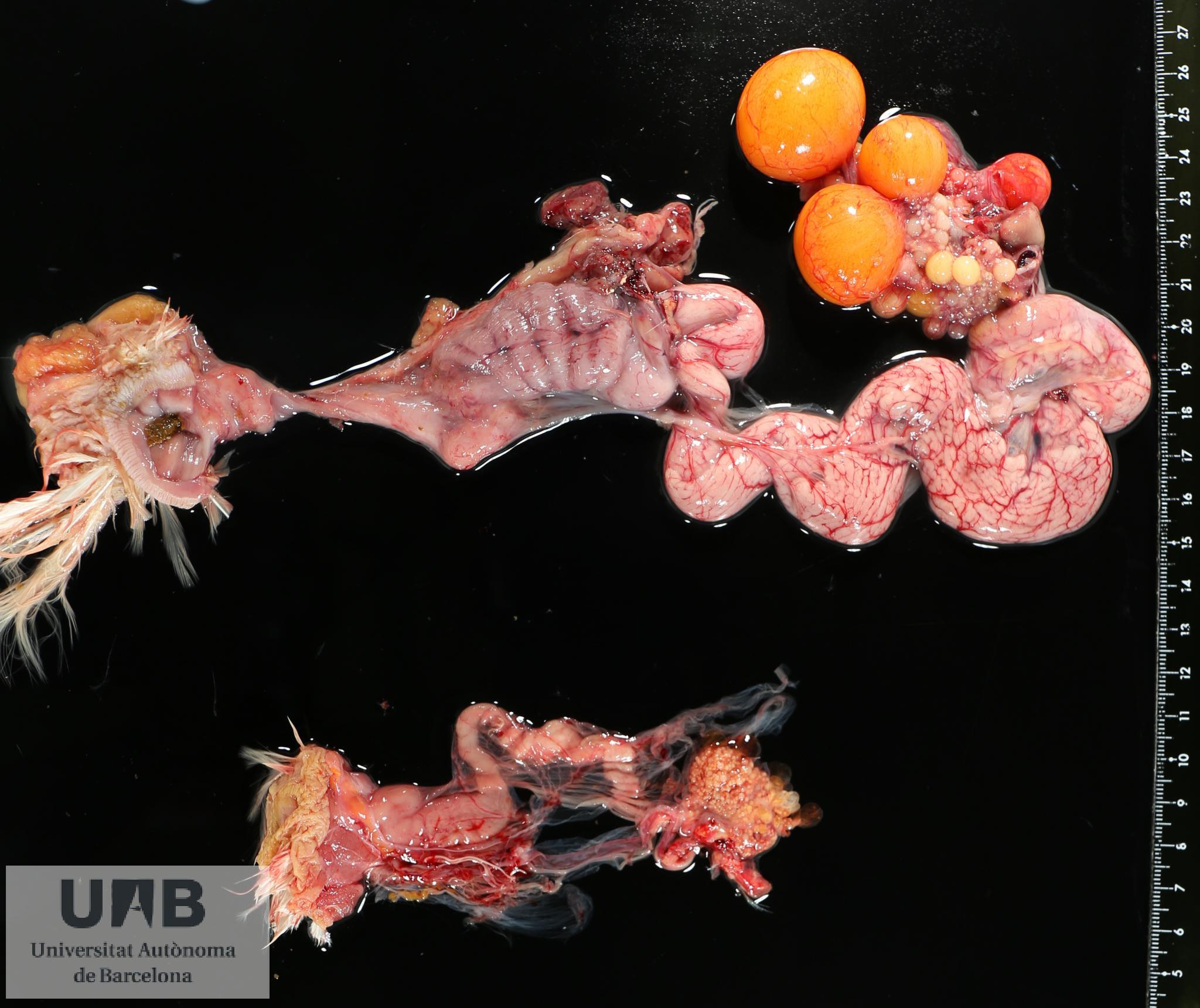
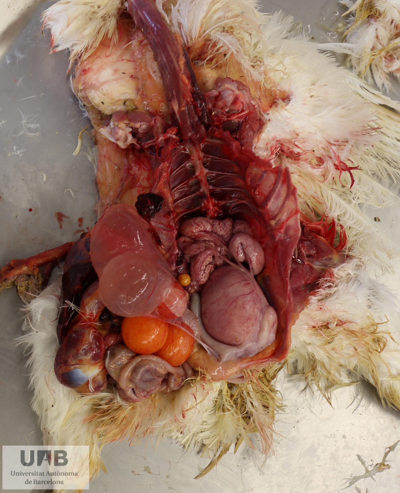
Laying hen carcass. In this case, when opening the coelomic cavity, a markedly dilated right oviduct could be seen, which accumulated watery fluid at its lumen: hydrosalpinx. As the right oviduct normally undergoes involution in chickens, the presence of persistent right oviduct is abnormal as well as the hydrosalpinx itself.
This finding is normally associated with avian bronchitis virus infection in laying hens. In the early stages of life, this virus is capable of replicating and harming the oviduct when it’s on its developmental stage, causing this degeneration of the oviduct and accumulation of fluid. Other common findings secondary to avian infectious bronchitis are left oviduct cysts, left oviduct atrophy and egg-yolk peritonitis.
In the image archive of the pathology department at the UAB Veterinary faculty there are good examples of these lesions:

1 comment(s)
Molt bona feina! Espero que en pugueu fer una altra edició aviat.