Any given day in a poultry slaughterhouse (2023)
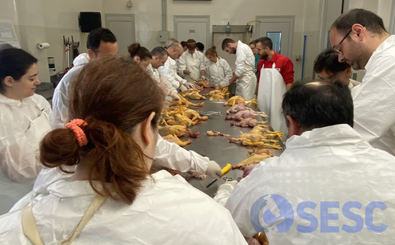
Last June we organized a practical workshop on the identification and description of lesions for official slaughterhouse veterinarians. For this reason, we collected viscera and carcasses condemned from several poultry slaughterhouses, which were the subject of discussion during the session.This post documents a couple of lesions from which samples were also collected in formalin for histological study. (AC)
The pathologists and collaborators of the SESC, Natàlia Majó and Carlos López-Figueroa, led the session.
CASE 1
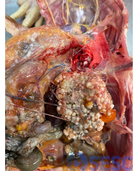
Laying hen carcasse. In this case, a large multinodular whitish mass is observed replacing the ovary. The serosas of the rest of the viscera have a rough appearance, with the presence of multiple miliary nodulations.
These macroscopic findings are characteristic of ovarian adenocarcinoma, the most common neoplasm in laying hens. This neoplasm usually prevents egg laying, and is a frequent finding in elderly laying hens.
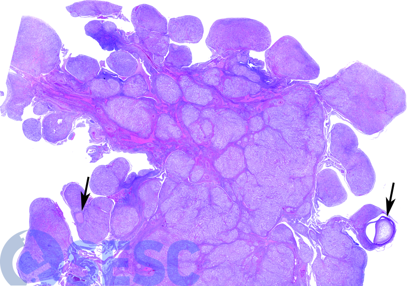
Histological section of the ovary. One can see how almost the entire ovary is replaced by an infiltrative multinodular neoplasm. Only two ovarian follicles are observed among the neoplasm (arrows).
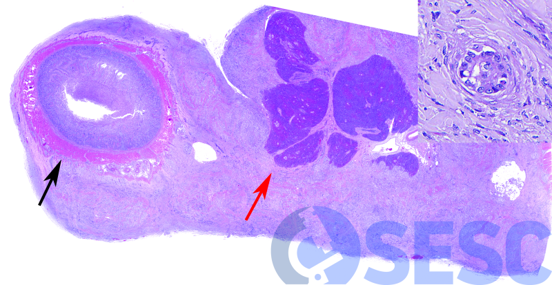
The histology of the duodenum-pancreas shows how this neoplasm has spread by seeding in the coelomic cavity (coelomic carcinomatosis), affecting the serosa of the intestine (black arrow) and the pancreas (red arrow). At higher magnifications (insert), neoplastic cells can be observed forming tubules and acini, of epithelial origin, confirming that it is an ovarian adenocarcinoma. In addition, very abundant mature fibrous tissue can also be seen between the neoplastic cells (fibrous desmoplasia), a common finding in these tumors.
CASE 2
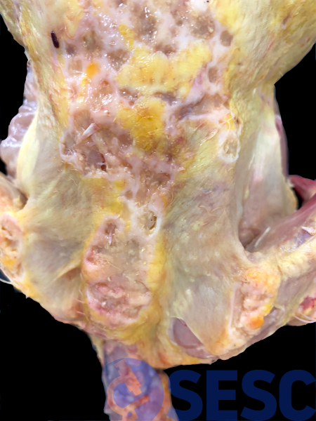
Laying hen caracass. In this case, multiple skin masses are observed on the back of the carcass. The skin in these regions is markedly thickened, in addition to having depressed, apparently ulcerated central regions (crateriform appearance).
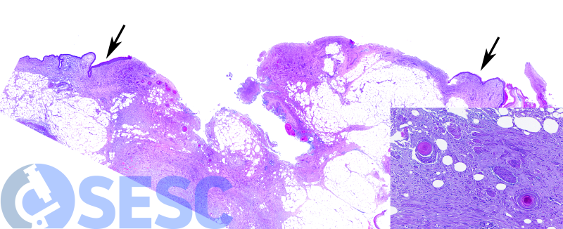
When carrying out the histopathological study, the presence of a skin neoplasm with central ulceration is confirmed. The presence of normal epidermis can be observed at the margins (arrows). The neoplasm is epithelial, and grows to form trabeculae and nests, which usually have eosinophilic (keratin-like) material inside.
These findings are compatible with squamous cell carcinoma, a malignant neoplasm of epidermal origin.
In this case it is necessary to remember the differential diagnosis of skin neoplasms in birds. The two most common are lymphoma associated with Marek's disease and squamous cell carcinoma. Marek's lymphoma tends to markedly affect the follicles of the feathers, and therefore initially appears as a thickening of these. On the other hand, squamous cell carcinoma has no predilection for any cutaneous structure, so as it grows it forms nodules and plaques irregularly distributed over the skin, which frequently ulcerate and take on the observed crateriform appearance.
