Myxomatosis in a batch of rabbits.
We received an inquiry regarding a batch of 300 rabbits in which 10 of them displayed lesions in the ante-mortem inspection, mainly regarding facial tumefaction, inflammation, and alopecia. The main suspicion of the official inspectors is myxomatosis. Three carcasses and facial skins from different animals were submitted to perform its anatomopathological study and eventually confirm or rule out whether it’s a case of myxomatosis.
Myxomatosis is a disease that affects lagomorphs (rabbits and occasionally hares) and it’s caused by viruses from the leporipoxvirus family (hence it’s a poxvirus). The disease is generally severe and has two distinct presentations: classic or cutaneous and respiratory or amyxomatous. Given its severity and repercussions in the industry and animal health implications, it is included in the list of mandatory declaration diseases of rabbits.
In the carcasses (without skin due to the slaughter procedures) the main finding was pulmonary cranioventral consolidation in all the carcasses, with different severity. At histopathological evaluation, a subacute interstitial pneumonia was diagnosed, which was mild in two carcasses and severe (with hyperplastic and necrotizing components) in the third. Also, mild suppurative tracheitis and rhinitis were observed. Myxomatosis can cause interstitial pneumonia in its respiratory presentation (with a proliferative – hyperplastic component), however in this case the histology was not fully conclusive.
However, the study of the skins revealed skin thickening and crusts in the regions of the ears, ocular conjunctiva, and snout. Additionally, the nares presented a dense yellowish exudate adhered to the skin and crusts of the snout. At its histopathological study, it consisted of a hyperplastic dermatitis associated with a myxoid proliferation of the dermis. Additionally, intracytoplasmic inclusion bodies characteristic of poxvirus were seen in the keratinocytes and fusiform cells in the dermis (classically named myxoma-cells). All these findings are compatible with the diagnosis of myxomatosis. Additionally, fungal structures compatible with dermatophytes were observed in the hair follicles of these skins (ringworm).
Lung and skin samples were submitted to the national reference laboratory for confirmation by means of molecular diagnostic. The presence of myxomatosis virus (classic strain) was confirmed by PCR in all the samples studied. Once again, the intervention of the veterinarian official inspectors was successful in diagnosing a disease of mandatory declaration such as myxomatosis. (AC)
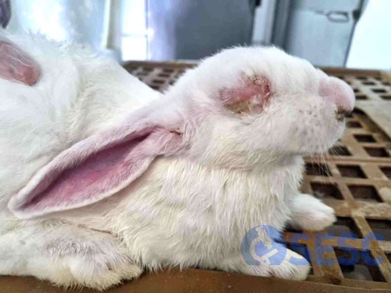
Rabbit that showed a marked tumefaction and thickening of the skin in the region of the ears, conjunctiva and snout. Additionally, yellowish dense exudate could be seen in the ocular conjunctiva and snout.
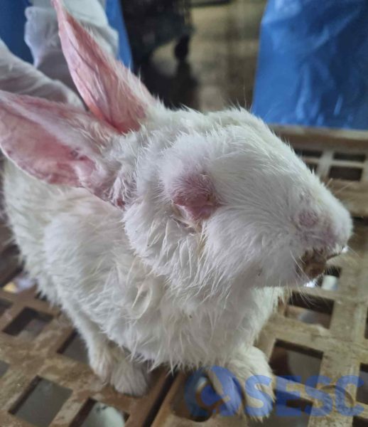
Rabbit with similar lesions, with general bad appearance of the hair and multifocal areas of skin thickening.
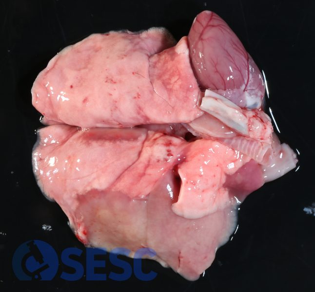
Extensive areas of cranioventral consolidation in the cardiac and diaphragmatic right lobes, displaying greyish coloration.
Proliferative dermatitis and conjunctivitis in one of the skins.
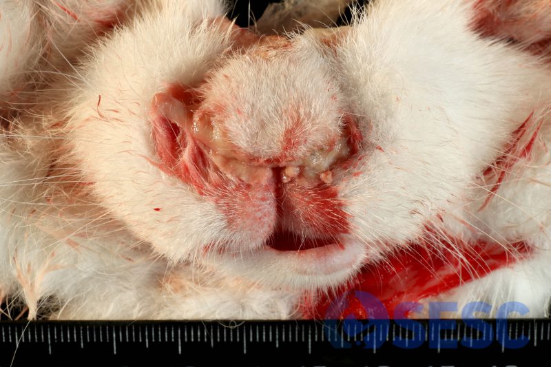
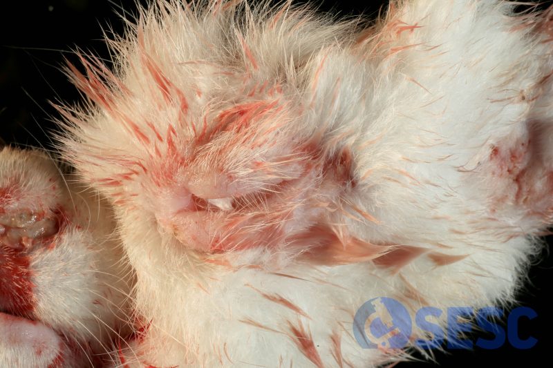
Similar lesions to those seen previously, in the snout region. Additionally, yellow dense exudate can be seen in the snout (suppurative rhinitis).
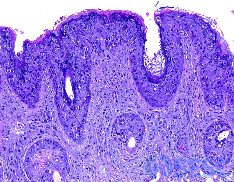
Histologic section of the skin, in which hyperplasia of the epidermis could be seen, accompanied by hydropic degeneration of the keratinocytes and abundant intraepithelial leukocytes (performing exocytosis). These histological changes also affected the follicular epithelium. The underlying dermis shows a mixed inflammation as well as proliferation of large fibroblasts, associated with a lax and slightly amphophilic matrix (myxoid).
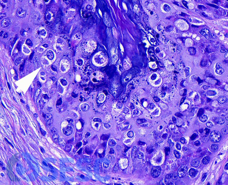
Detail of the histologic changes in the epidermis. Occasional intracytoplasmic inclusion bodies could be seen in the keratinocytes (arrow).
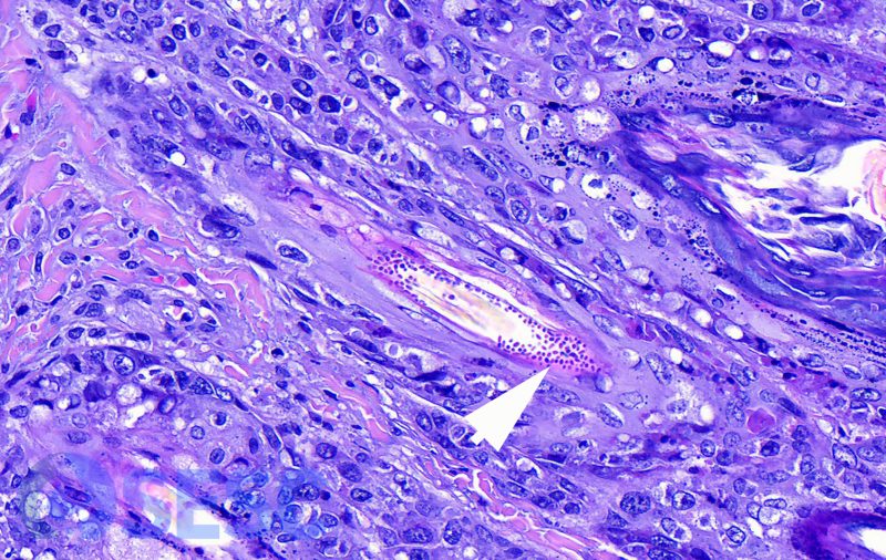
Skin section in which all the above-mentioned changes can be seen. Additionally, fungal rounded structures can be seen in the periphery of a hair in its follicle, compatible with dermatophytes (arrow).
