Chronic suppurative bronchopneumonia with bronchiectasis in cattle
From time to time, the SESC receives inquiries to rule out tuberculosis in animals that arrive at the slaughterhouse with lesions of chronic suppurative bronchopneumonia with bronchiectasis (dilation of bronchioles due to the presence of purulent or suppurative exudate inside them). These lesions are relatively common and are associated with bacterial infections: we have often isolated Mycoplasma bovis in combination with other bacteria such as Pasteurella multocida or Corynebacterium sp.
In this post we have tried to briefly define some criteria to differentiate this type of chronic lesion from tuberculous lesions since, macroscopically, they can present some similarities. The post also includes images of several examples to familiarize you with this lesional pattern.
In case of doubt, however, it is always necessary to submit samples for a laboratory study to rule out that it is tuberculosis, especially now that Catalonia has been officially declared free of bovine tuberculosis and it is more relevant than ever to be able to make an early diagnosis of potential new outbreaks. (EV)
Table 1: Criteria for differentiating chronic suppurative bronchopneumonia from tuberculous pneumonia.
| Chronic suppurative bronchopneumonia with bronchiectasis | Tuberculous pneumonia | |
| Distribution of the lesions | Usually follows a cranioventral distribution (involving the apical and middle lobes and the cranioventral part of the diaphragmatic lobe). | Focal or multifocal lesions, of variable size and distribution. |
| Appearance of the lesions | Often has a characteristic multinodular appearance. The nodules correspond to the dilated bronchi full of suppurative exudate, which is rather fluid and yellowish to greenish in color (nodules may be a little hardened as they may present a certain degree of mineralization and fibrosis). | Granulomatous lesions with a more solid , caseous, texture, yellowish to whitish in color, with a necrotic component, which is usually mineralized (hardened and crackling when cut), they can also have some degree of fibrosis.
Tuberculosis can also cause multinodular pearly lesions in serous membranes such as the pleura. |
| Lesions in the lymph nodes (tracheobronchial and mediastinal) | This type of lesion is usually associated with lymphadenomegaly due to hyperplasia of the respiratory lymph nodes, but without the conspicuous macroscopic presence of pus or caseum. | Tuberculosis usually causes granulomatous lesions in the lymph nodes. |
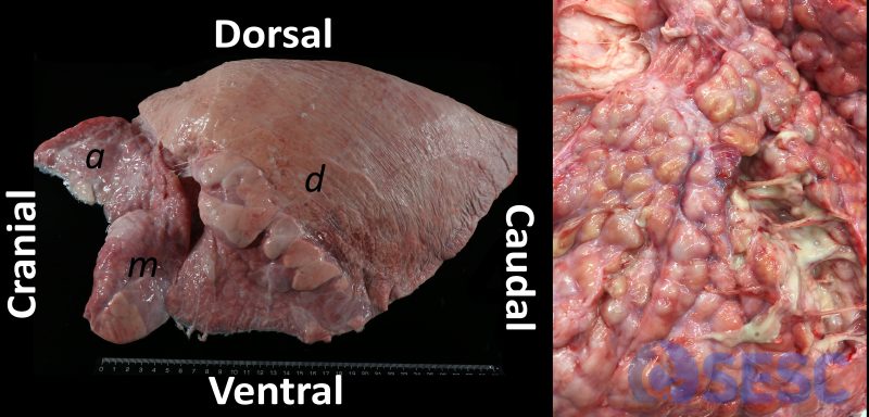
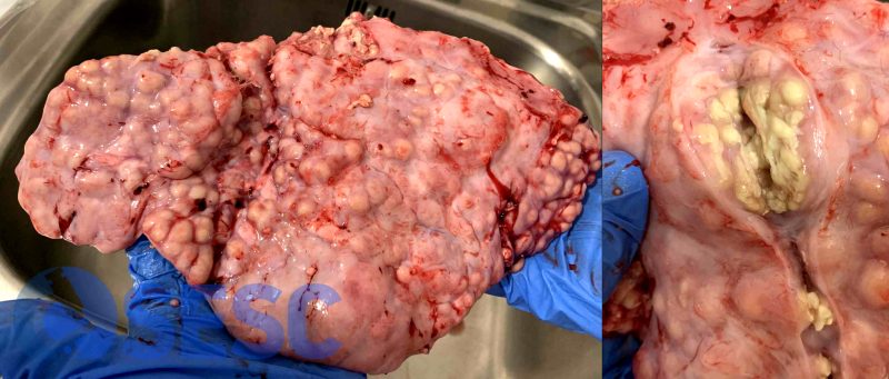
Paradigmatic example of chronic suppurative bronchopneumonia with bronchiectasis in a calf lung with the typical cranioventral distribution pattern. The different lung lobes are indicated: a. Apical; m: middle and d: diaphragmatic. On the right, a clser look at an example of the appearance of bronchiectasis, its purulent content can be seen in the sectioned ones.
In this case referred to the SESC, Mycoplasma bovis and Corynebacterium sp. were isolated.
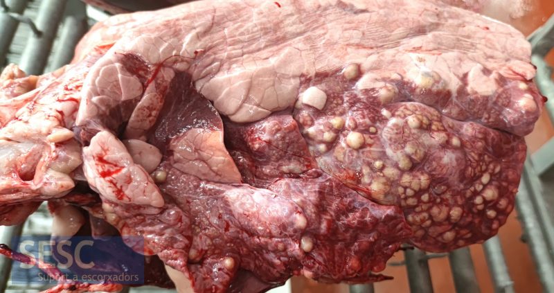
En aquest altre cas es va aïllar Mycoplasma bovis, Pasteurella multocida i Corynebacterium sp.
In this case, Mycoplasma bovis and Pasteurella multocida were isolated. You can read the full case here.
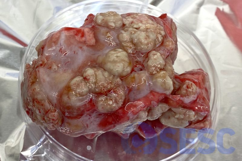
Characteristic multinodular lesion. Once secitioned, its appearance might be similar to a granulomatous lesion, as it can also present some degree of mineralization.
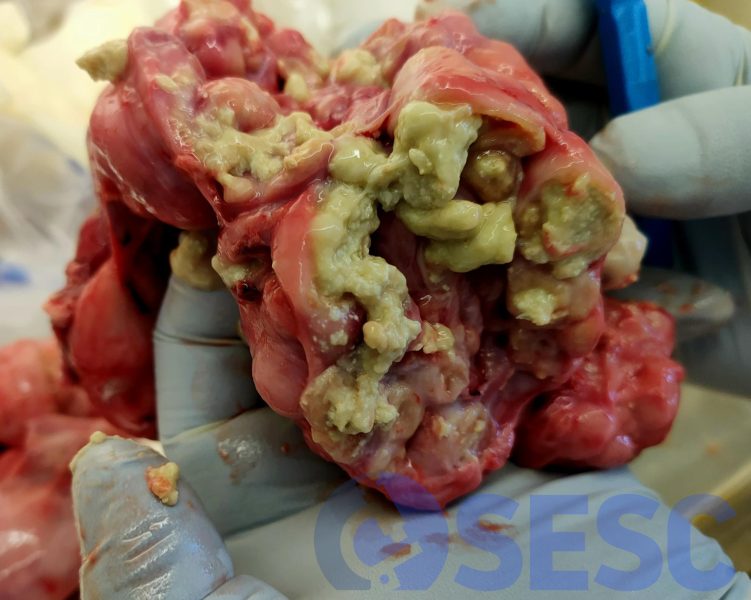
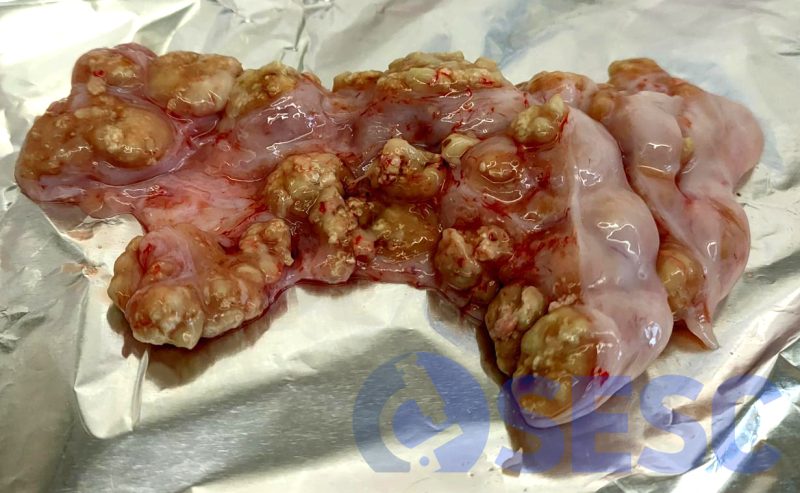
Once sectioned, its appearance might be similar to a granulomatous lesion, as it can also present some degree of mineralization, but a more fluid suppurative component can also be observed.
