24/07/2023
|
Abdominal cavity (porcine)
0
What is your diagnosis? (117)
At the end of the post you can vote for the right diagnosis!
Click on the option that you think is correct and then click on the “Submit” button to see if you’ve succeeded.
A 6-month-old female hybrid breed pig carcass shows multifocal nodular lesions in the abdominal wall and liver, 3 to 5 cm in diameter, with a fibrous wall, and cream-colored pasty/fluid content. (EV)
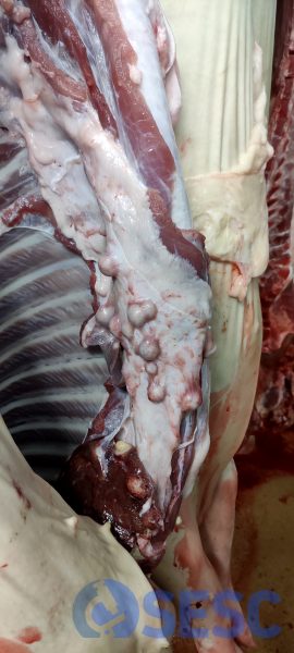
Multifocal nodular lesions adhering to the serosa on the abdominal side of the diaphragm.
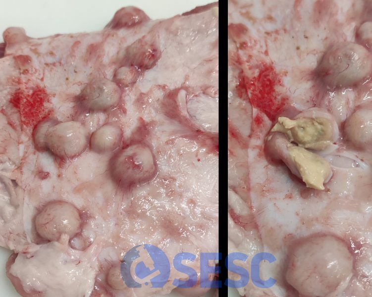
Detail of the lesions, on the right you can see the contents of one of the nodules upon its section.
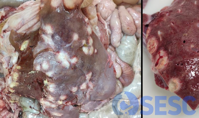
The lesions are also localized in the liver parenchyma, with a generalized multifocal distribution.