Coelomic parasitosis by Ascaridia galli and uterine leiomyoma
An inquiry was submitted about a laying hen of unknown age, which was cachectic and, on the post-mortem inspection, two findings were detected in the coelomic cavity. First, a mass of firm consistency was seen adhering to the serosa of the oviduct. The other finding consisted of a free nematode parasite in the coelomic cavity. The official meat inspectors requested a diagnosis of the tumor and identification of the parasite.
We received the entire carcass and proceeded to necropsy it. The tumor, located in the serosa of the oviduct but independent of the muscular wall, had a large size and a firm consistency, and on section it had a slightly variegated coloration. The parasite was collected from the coelomic cavity and, upon opening the intestine, abundant nematodes with similar characteristics were observed, which were also collected.
The histological study of the tumor revealed a uterine leiomyoma, a benign neoplasm originating most likely in the muscle cells of the wall of the oviduct. On the other hand, all the parasites collected both in the coelomic cavity and in the intestine were sent to the parasitology department for their identification, and it was concluded that all the adult nematodes corresponded to specimens of the species Ascaridia galli. It is known that these nematodes can migrate from the intestine and through the cloaca to the oviduct, appearing in its lumen, inside eggs, or even reaching the coelomic cavity. The presence of specimens in the intestine is consistent with this fact. The presence of coelomic parasitism and the tumor in the same animal are independent and coincidental facts. (AC)
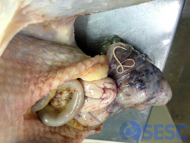
Image from slaughterhouse in which the parasite is observed on the tumor, in the coelomic cavity.
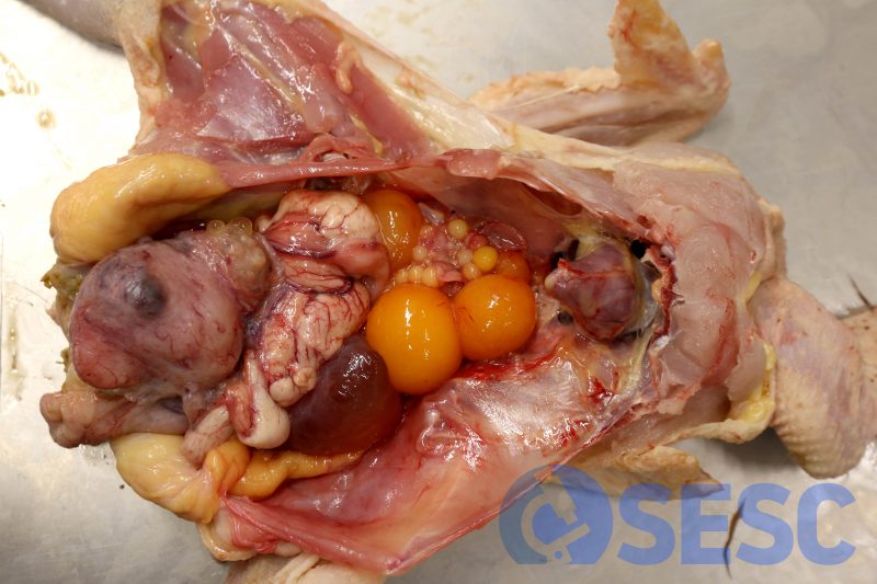
Opening of the coelomic cavity during necropsy. The mass attached to the reproductive system is appreciated.
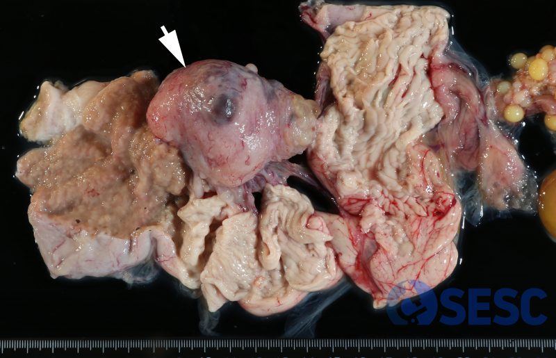
Once the reproductive tract is removed, it could be seen how the tumor (arrow) is attached to the serosa of the oviduct but not to the muscular wall.
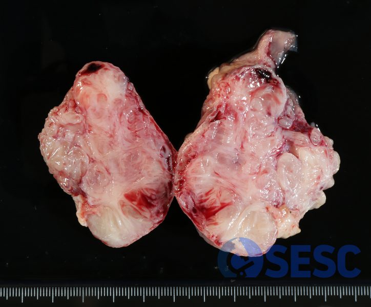
Section of the tumor in which the fibrous appearane is observed, finely variegated.
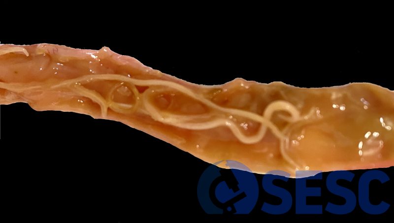
Presence of nematodes in the intestinal lumen.
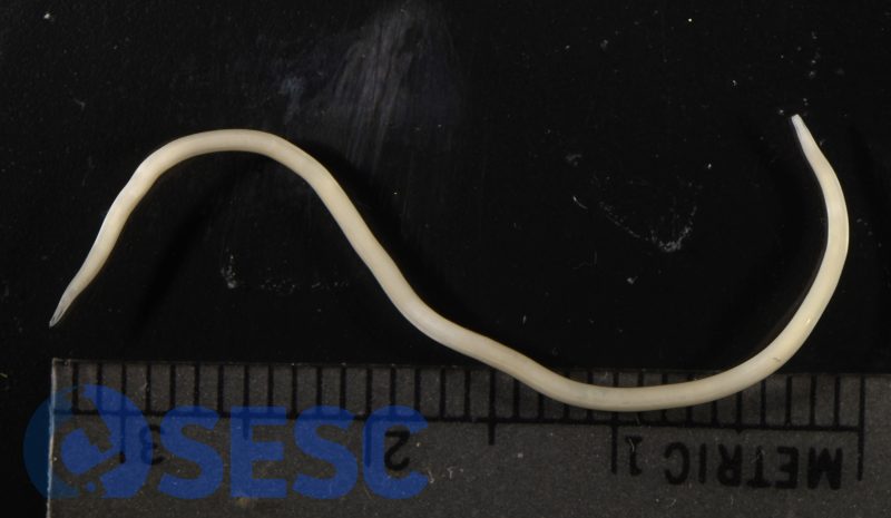
Female Ascaridia galli extracted from the intestine. Parasitic diseases, Faculty of Veterinary Medicine UAB.
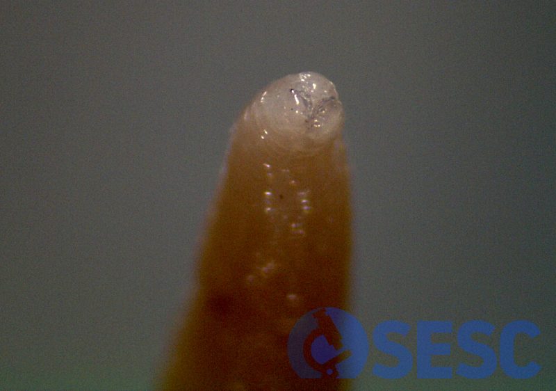
Magnifyed image of the parasite, where the lips can be observed, which are one of the features used to identify the species. Parasitic diseases, Faculty of Veterinary Medicine UAB.
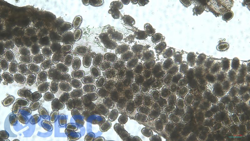
Eggs in the uterus of the female Ascaridia galli. Parasitic diseases, Faculty of Veterinary Medicine UAB.
