02/12/2013
|
Poultry
2
Fungal dematitis in a chicken carcass
The plaques were soft, of increased skin thickness and, when cut, were yellowish in color and edematous with the presence of small nodules of 2-3 mm of diameter. Histopathological study showed granulomatous inflammatory lesions. Grocott staining to identify the presence of hyphae in the center of the granuloma was performed with a positive result. A diagnosis of fungal granulomatous dermatitis was established.
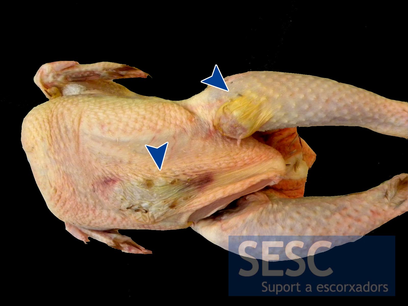
Thickened plaques (arrows) in the skin of an organic chicken carcass.
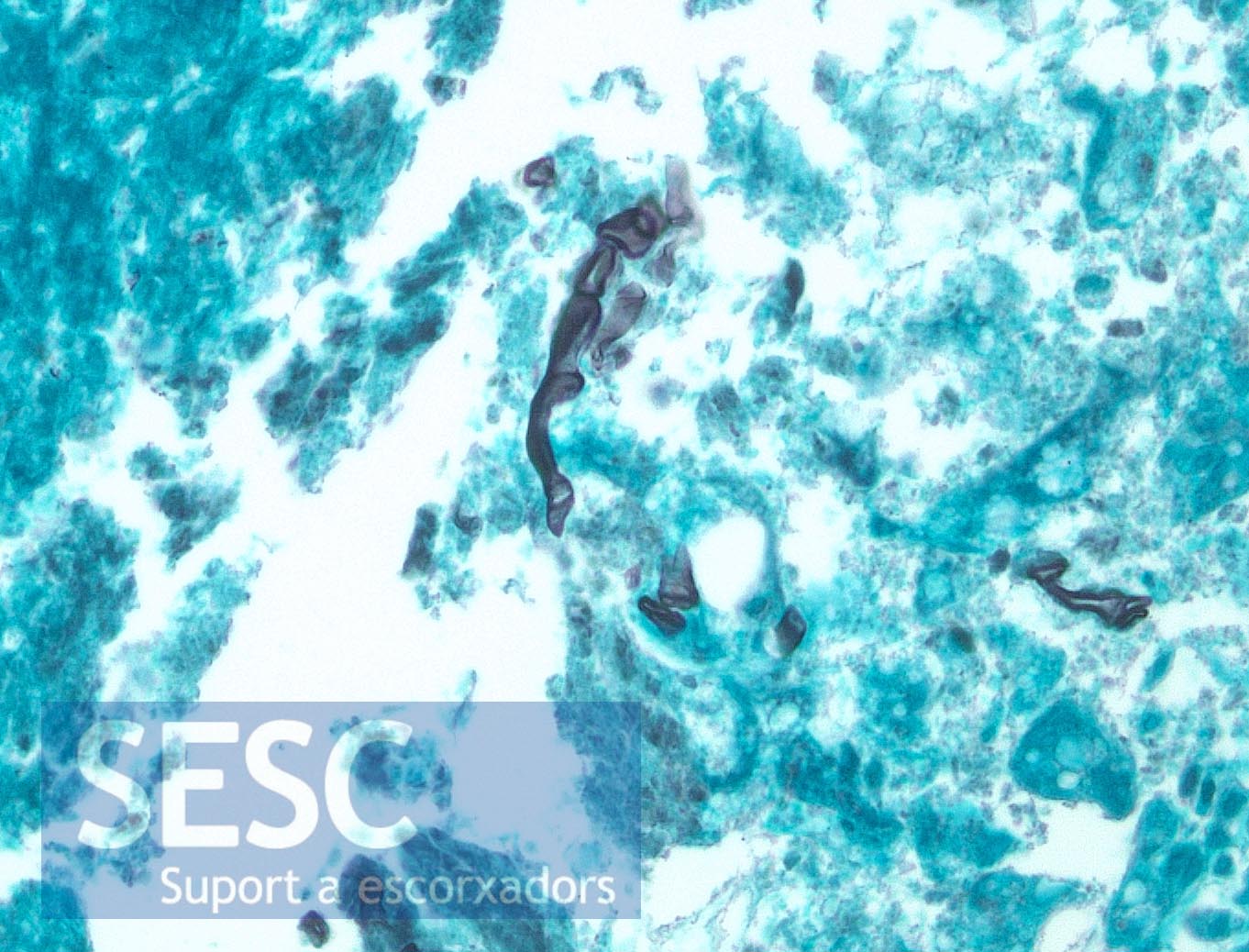
Grocott staining evidencing the hyphae (black).

2 comment(s)
Comment from Veterinary Pathology group in LinedIn:
D.A.: Have you set up fungal cultures from cases like this, and if so, what did you grow?
Our pathologist answer: Unfortunately we did not process any samples for fungal culture.
It is the first time we diagnose this in a chicken, “organic chicken” pathology is an emerging field here, so we cannot give you previous results. However the lesion type is suggestive of a Zygomicoses.
Comment from Livestock Health group in LinkedIn:
O.P.: My experience includes various predisposing factors especially in tropical poultry production ranging from poor husbandry, unfavorable ambient conditions to complicate initial prediposition to the pathogenic fungi organisms. This is a common complication in broiler chickens raised under poorly ventilated pens with wet floor. In addition, there would be infectious foot-pad dermatitis and chronic gangrenous inflammations of the breast tissues