Lesions of Epizootic Hemorrhagic Disease in cattle
Epizootic Hemorrhagic Disease (EHD) has been confirmed for the first time in Spain in two cattle farms in the provinces of Cádiz and Seville, in addition to suspicions of compatible clinical symptoms in 25 other farms so far. Spain has lost its EHD-free status. Updated information regarding outbreak monitoring can be found in the agricultural ministry's official sources. From SESC, we want to take the opportunity to revisit EHD from a pathological point of view, and discuss the lesions that could be observed at the slaughterhouse.
EHD is a disease transmitted by the bite of a biting midge (Culicoides), caused by an orbivirus close to the one causing bluetongue in small ruminants. In fact, the disease it causes is comparable to bluetongue, although it is generally less severe. It infects and causes disease in cattle, which is rarely serious or fatal. Infection in wild ungulates such as deer is generally more severe (estimated to have mortality rates of 20% or greater). Sheep and goats can be infected with the EHD virus but rarely develop clinical signs.
EHD virus infection results in endothelial replication and vascular damage, which is primarily responsible for the clinical signs and lesions seen in infected animals. Clinical signs include fever, facial (primarily conjunctival) edema, ocular and/or nasal discharge, salivation, dyspnea, erythema or desquamation of the snout, lameness, and udder erythema.
With regard to the lesions that could be seen in the slaughterhouse, it must be considered that in many cases they can be mild, not very specific, and that it must be supplemented with the ante-mortem examination. The lesions are most frequently confined to the oral cavity and consist of congestion, petechiae, hemorrhages, vesicles, erosions and/or multifocal ulcerations of the oral cavity, tongue and/or lips or coronal region (transition between skin and hoof). The most severe form, known in Japan as Ibaraki's disease (a form of MHE caused by serotype 2), is more similar to bluetongue in sheep, with edema and hemorrhages in other locations. The differential diagnosis must include foot-and-mouth disease, bovine viral diarrhoea, infectious bovine rhinotracheitis, vesicular stomatitis, malignant catarrhal fever and bovine ephemeral fever.
In the case of finding suspicious cases, it is important to send samples to the Central Veterinary Laboratory (LCV, Algete) to rule out the suspicion by means of a PCR since the EHD is subject to official control. The samples of choice are: a swab of vesicular/ulcerated lesions (ante-mortem and post-mortem) or spleen, lymph node and lung (post-mortem). (AC)
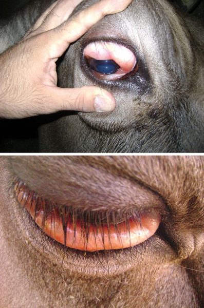
Conjunctival edema in a cow. Source: CDC-EID.
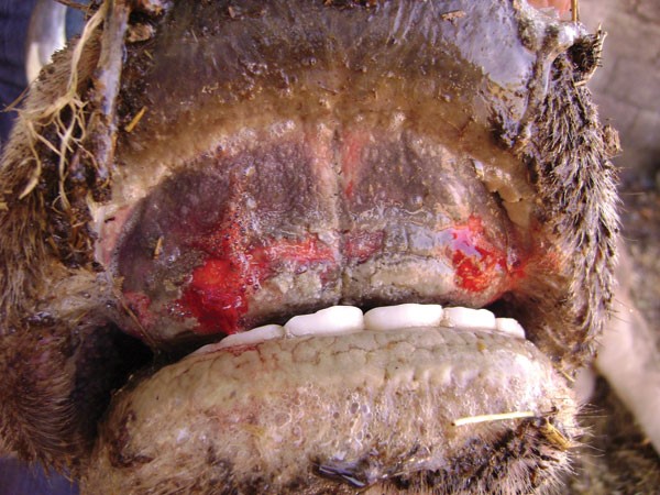
Cattle. Ulcerative lesions in the oral mucosa. Source: CDC-EID.
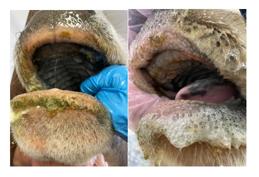
Cattle with hypersalivation and ulcerative lesions on the oral mucosa. Source: Cornell University, College of Veterinary Medicine.
