The curious case of the sheep with “green” lymph nodes
We received an inquiry about an ovine carcass that shows a greenish coloration of all the lymph nodes (preescapular, inguinal, renal...). At post-mortem examination, no further macroscopic alterations within the remaining viscera were recorded. We received a sample of one of the affected lymph nodes, in order to perform its histopathological assessment and try to elucidate the nature of this lesion.
Histologically, a profound infiltration of mature polymorphonuclear eosinophils was seen in the lymph node, with multifocal to generalized distribution and basically associated with the lymphatic sinuses of the organ, surrounding the entire pre-existent lymphatic tissue (lymphatic follicles). Additionally, diffusely, the same eosinophil population infiltrated the capsule and the pericapsular tissue. The lymphoid tissue presented a mild activation and hyperplasia.
The histologic findings were readily characterized, and in fact, are consistent with the macroscopic appearance of the lymph nodes. The presence of high numbers of eosinophils gives tissues a greenish coloration, similarly to what happens in bovines with eosinophilic myositis, which shows an evident greenish coloration. However, the diagnosis and the aetiology of this process remained on discussion. The following options were considered:
- Parasitic: the parasitic reactions usually are eosinophilic, and accumulation of eosinophils in the regional lymph nodes can be expected. However, in this case it’s improbable due to all lymph nodes showing the same change. Additionally, such massive infiltrations in the lymph node are not described as an answer to parasites.
- Myeloproliferative disorder: in humans the chronic eosinophilic leukemia and the hypereosinophilic syndrome are recognized. Both correspond to a systemic proliferation of mature eosinophils that are released into the circulation and can variably infiltrate other organs. The leukemia is a neoplastic process and the hypereosinophilic syndrome is its reactive counterpart, however the line between the two conditions is often blurry. Despite not being formerly described in sheep and without knowing the age of the animal, this option was considered as a possibility. To help confirm or rule out this possibility, it would have helped to assess other organs such as bone marrow, liver and spleen, organs which typically present myelopoietic cells in proliferative context (both neoplastic and reactive).
- Mast cell tumour: the option that the eosinophilic infiltration was secondary to a disseminated mast cell tumour was considered, a relationship frequently, observed. To investigate this, an immunohistochemical study against C-Kit was performed, and an increased presence of mast cells relative to a healthy lymph node from a healthy sheep was not observed.
- Similarly, type I hypersensitivity reaction (IgE mediated) typically features an intense recruitment of eosinophils which is mast cell mediated.
Unfortunately, in this case a conclusive diagnosis was not achieved even though the pre-existent bibliography was deeply checked. However, these cases often result the most interesting ones as they show a great opportunity for discussion and revision. Maybe this mystery will remain unanswered or future cases will help to investigate further. The SESC team would welcome your feedback, and if you have ever come across “green” lymph nodes, you could leave a comment. (AC)
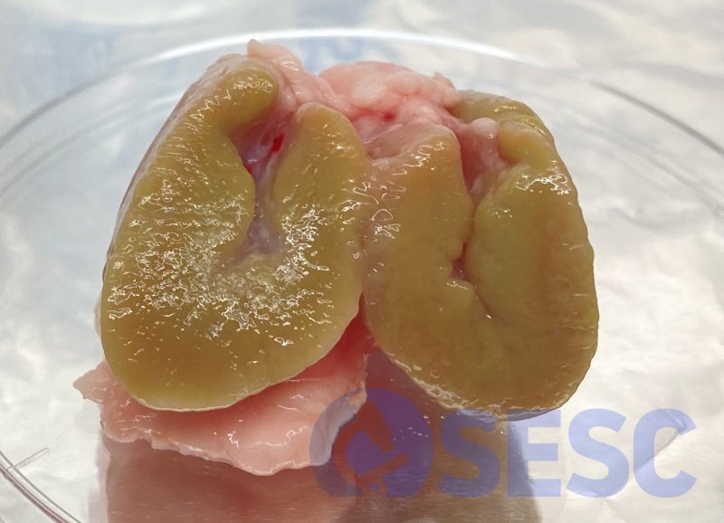
Lymph node of a sheep carcass. At the cut section, a diffuse greenish coloration can be seen in the entire lymph node.
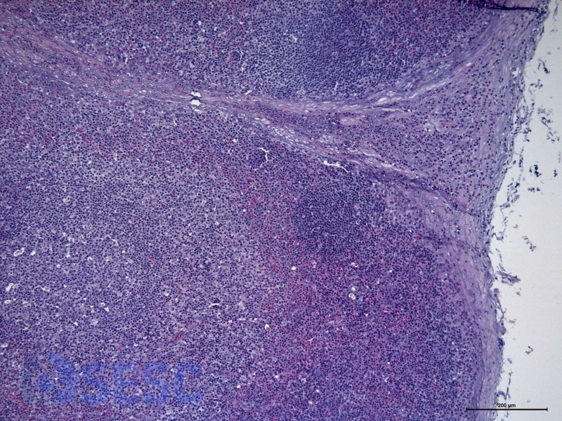
Presence of eosinophils in the subcapsular sinuses, surrounding the lymphoid follicles.
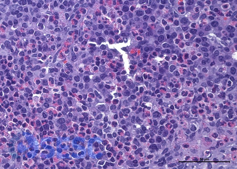
Detail at higher magnifications of the eosinophils infiltrating the lymph node. The eosinophils appear well differentiated, judging from its presence of conspicuous reddish granules and binucleated nuclei.

2 comment(s)
Sou inspetora sanitária num matadouro na zona da cidade de Tomar, Portugal, e encontro esta situação com bastante frequência. Como normalmente os ovinos geralmente estão parasitados, sempre associei esta coloração esverdeada à presença de eosinófilos secundários ao parasitismo.
Talvez seja um caso de CHLORELOSE.