The curious case of the orange chickens.
We received an inquiry regarding three laying hens that were condemned for showing a bright intense orange coloration of the skin. We received two of them for diagnosis, so their necropsy was performed. During the necropsy, the laying hens presented signs of virilization (comb and wattle overgrowth, increase in spur size…), as well as a very intense orange coloration of the skin and the subcutaneous and perivisceral adipose tissue. We performed the alcohol-ether determination, and it was confirmed that this coloration was due to an accumulation of carotenoid pigments.
Furthermore, during the necropsy ovarian tumors were found in both of the animals, one of them presenting also masses in the intestinal serosa (metastatic seeding). The histologic study of this masses was performed and they were diagnosed as arrhenoblastomas. This tumor is classified as a sex cord-stromal tumor, and is characterized by producing virilization due to the production of androgens or inhibin. In the case of avian species, usually structures resembling seminiferous tubules can be seen, even with spermatogenesis.
Although these tumors explain the virilization of the laying hens, the cause of the hyperpigmentation dindn’t seem clear at a first glance (remember that this was the cause of the condemnation). One would expect that all the animals within a batch eat the same feed, thus the same quantity of carotenoids. However, laying hens usually deposit large amounts of the ingested pigments within the egg yolk, which gives it its orange coloration. As this hens had ovarian tumors and masculinization, they were not producing eggs, and thus the lack of pigment deposition within the yolk was likely causing its accumulation within the adipose tissue. (AC)
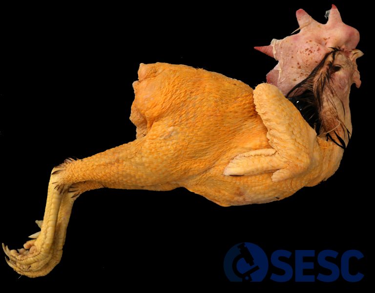
Laying hen with very intense orange coloration of the skin and masculinization (large size of comb and wattles and spurs).
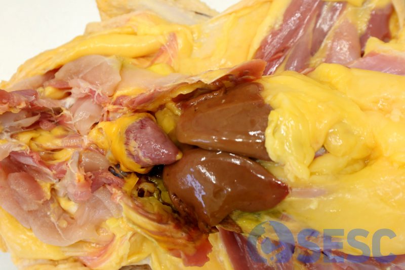
When the skin is removed and the coelomic cavity is opened, a very intense orange coloration of the subcutaneous and perivisceral fat is observed (especially noticeable in the epicardial fat).
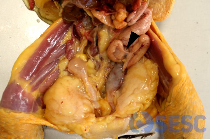
Coelomic cavity of one of the hens, in which a solid mass can be seen replacing the ovary (arrow). A left hydrosalpinx is also observed.
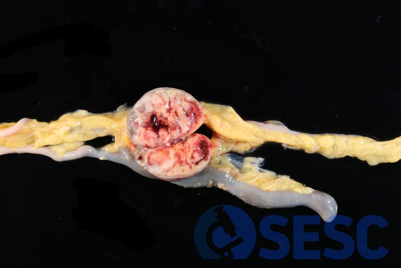
Mass adhered to the intestinal serosa (seeding metastases).
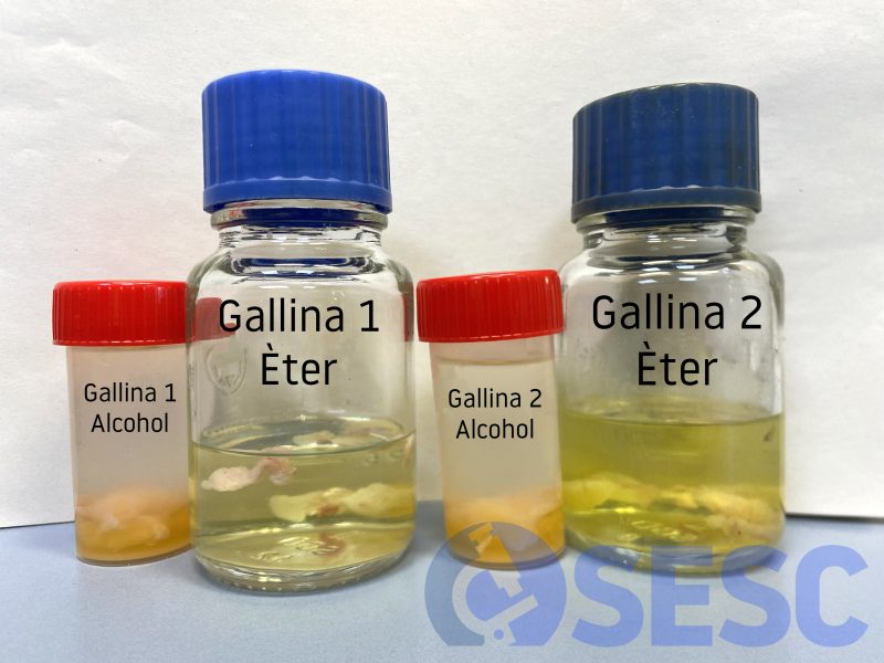
Alcohol-ether test. When adipose tissue is placed in alcohol, it does not change color. On the other hand, the ether turns to an intense yellow-orange colour. This confirms that what gives color to adipose tissue are carotenoid pigments, because these are soluble in ether and not in alcohol. You can read more about this determination in this post.
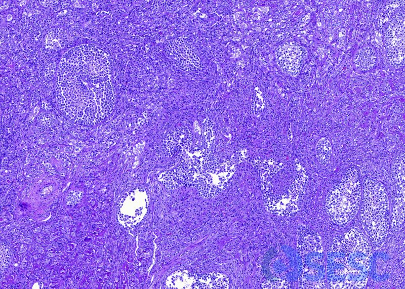
Histological detail of arrhenoblastoma. A heterogeneous neoplasm can be observed given the diverse origin of the cells that make it up.
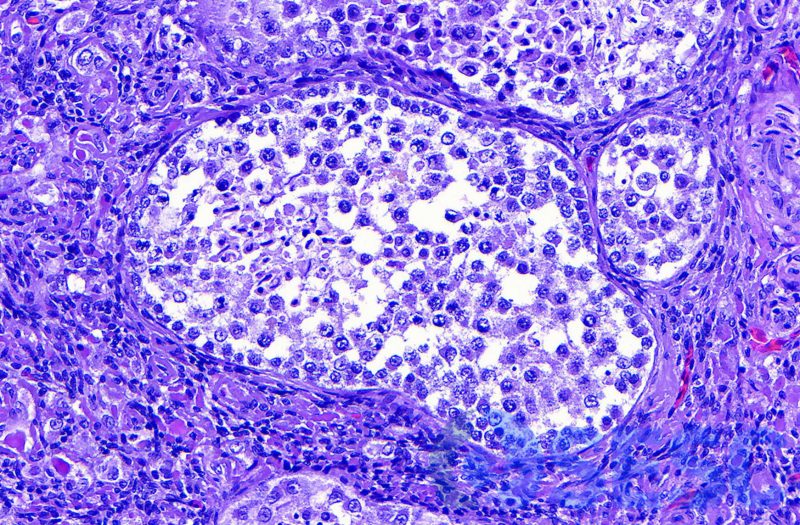
Histological detail of an intraneoplastic structure reminiscent of a seminiferous tubule. The elongated and pyknotic nuclei inside the tubule indicate a certain degree of spermatogenesis.
