Any given day in a pig slaughterhouse (2023)
Last June we organized a practical workshop on the identification and description of lesions for official meat inspectors. For this reason, we collected viscera that were declared not apt for human consumption in two swine slaughterhouses, which were the subject of discussion during this session. In this post some of the lesions that were observed are collected, as well as its histological study. (AC)
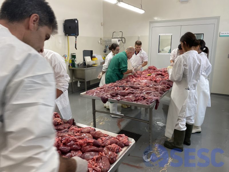
Pig expert pathologist Quim Segalés (IRTA-CReSA, UAB) participated in the discussion of the lesions observed in viscera declared unfit.
Case 1
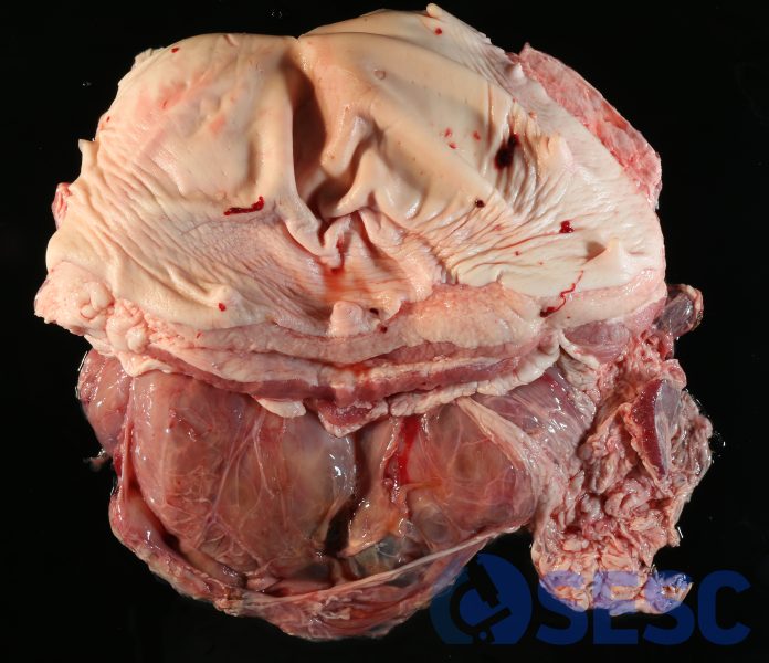
Skin and abdominal wall. One of the condemned viscera consisted in abdominal wall (including skin, abdominal musculature, and peritoneal parietal surface). It was attached to a portion of spleen by its inner surface. It presented a big, localized tumefaction, which fluctuated upon palpation.
When cut, it consisted in big cystic cavities delimited by bands of fibrous tissue and full of serous fluid.
This finding is compatible with an hygroma of the abdominal wall. It is a very chronic lesion and without much clinical significance. These lesions tend to appear when animals continuously rub parts of their bodies, generating a sterile chronic inflammatory lesion with fibroplasia.
Case 2
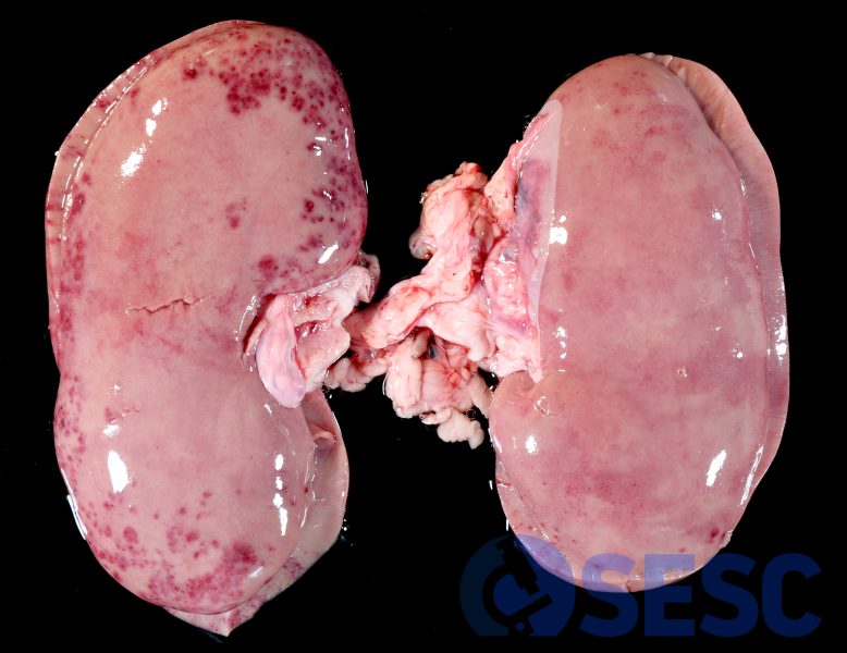
Porcine kidneys. In the cortical surface of the kidneys, multifocal to coalescent reddish areas can be seen. Remarkably, the distribution of the lesions within the renal poles is very suggestive of pyelonephritis.
A pyelonephritis is the ascendent inflammation (from the urinary tract) of the kidneys by bacteria. The papillae in the poles of the kidneys of the pig (due to anatomical conditions) are much more susceptible to urine reflux and hence, bacteria usually gain access to the renal tubules easier in these regions.
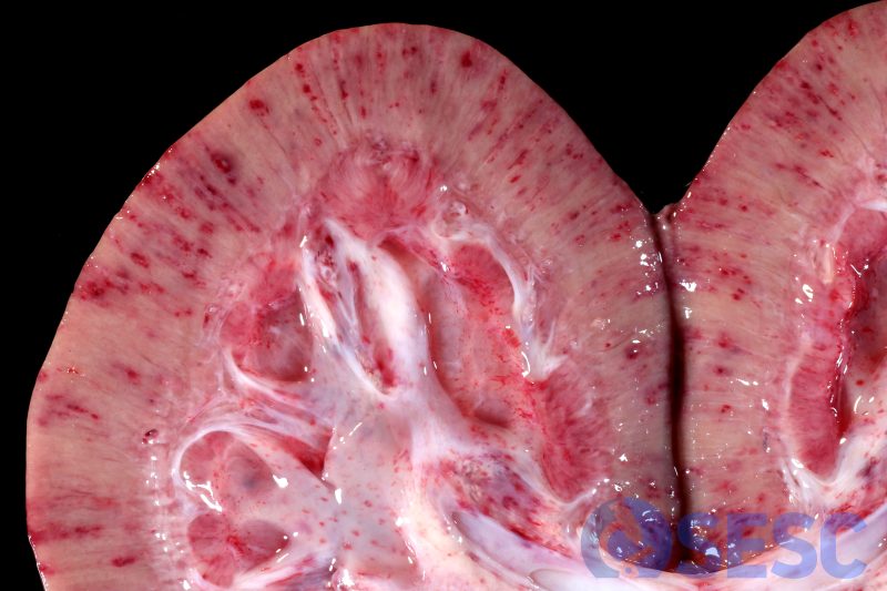
When sectioned, these foci could be observed as reddish areas in the cortex with radial pattern. Petechia in the renal pelvis could be also observed.
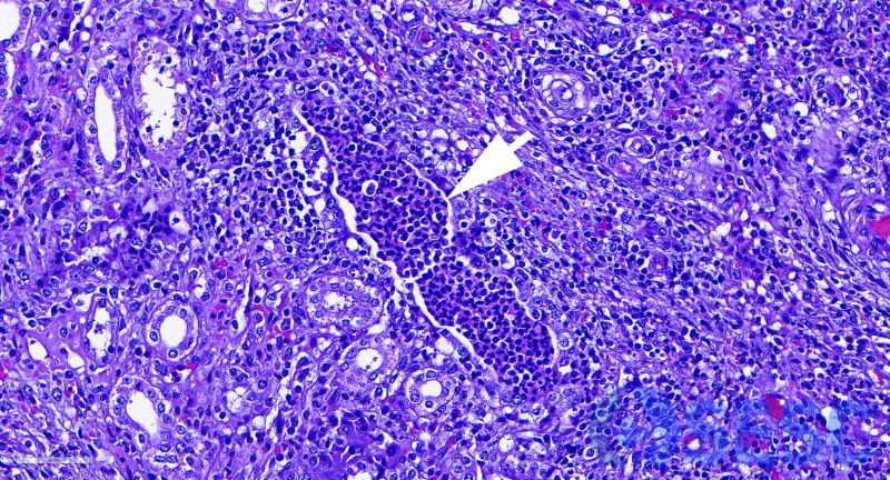
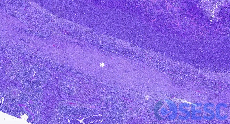
Histologically it was confirmed the presence of pyelonephritis, observing big accumulations of neutrophils within the lumen of renal tubules (arrow).
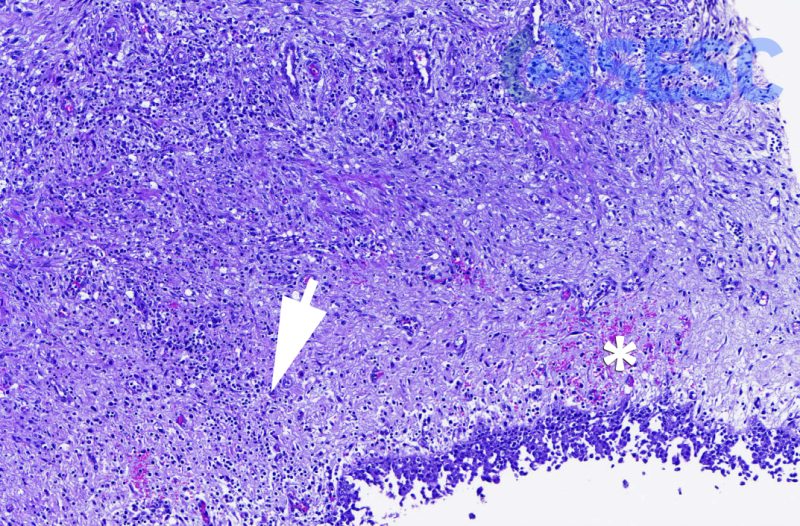
Chronic inflammation in the pelvis (arrow) and microhemorrhages (asterisk) were also observed, which were also macroscopically evident.
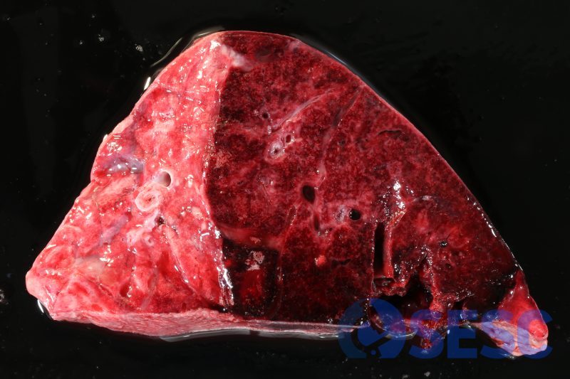
Case 3
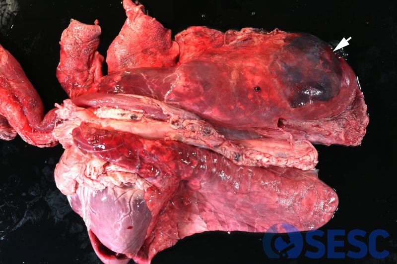
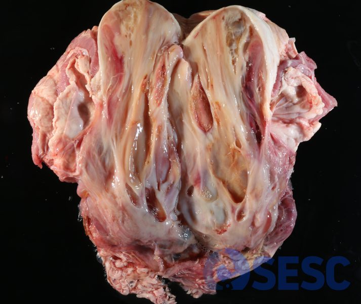
Porcine lungs. In the right diaphragmatic lobe of the lungs, a well circumscribed lesion could be seen, which was prominent, consolidated and with a reddish coloration (haemorrhagic and probably necrotic), with fibrin clots on the pleural surface (arrow).
Cut section of the affected lung.
All these features are characteristic of a fibrino-hemorrhagic and necrotizing pleuropneumonia. Usually, Actinobacillus pleuropneumoniae and other high pathogenicity bacteria are the causal agents, and are characterized by a suppurative bronchopneumonia in which a haemorrhagic and necrotizing component is overimposed due to the generated toxins.
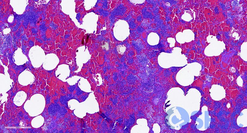
Histologically necrosis, haemorrhage and abundant exudation of fibrin can be observed.
Case 4
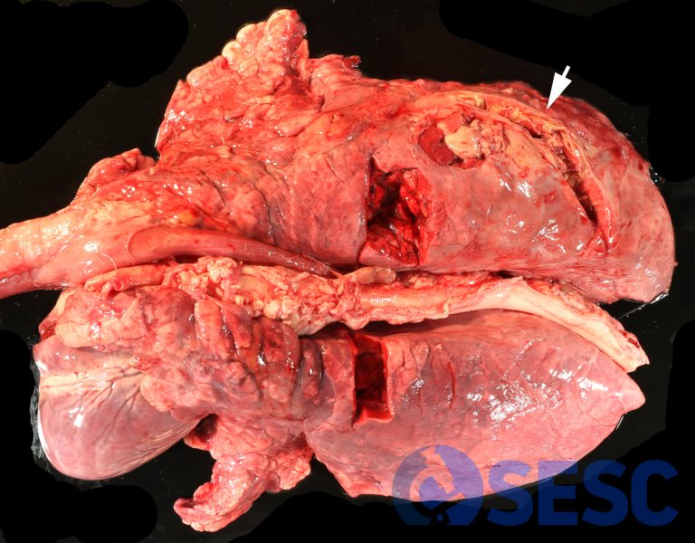
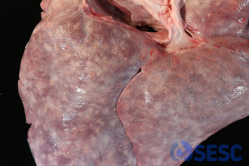
Porcine lungs. In this case a lesion was observed which was similar in distribution. However, this lesion presented a rather whitish coloration, and was hardened upon palpation.
At the cut section, big accumulations of purulent to caseous content can be observed in a irregular pattern.
This lesion is the consequence of the cronification of the anterior lesion (fibrino-necrotizing pleuropneumonia). If the animal survives the acute phase (occasionally associated to antibiotic treatment or previous vaccination), the pulmonary tissue often undergoes coagulative necrosis, and a thick fibrous capsule is generated with the intention of circumscribing the necrotic lesion. This phenomenon is known as pulmonary sequestration.
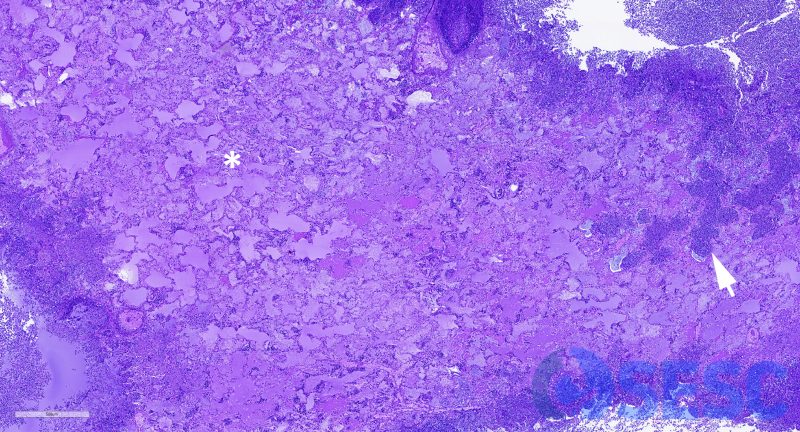
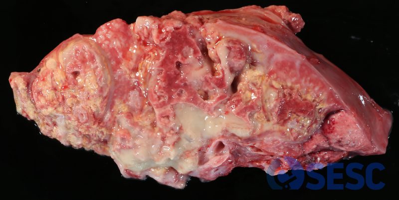
Histologic detail of this lesion. Abundant areas of coagulative necrosis can be observed (alveolar structure is still evident, asterisk) as well as dense areas of inflammatory infiltrate (arrow).
The necrotic tissue is delimited by a thick capsule of mature fibrous tissue (asterisk), being the consequence of viable tissue demarcating the necrotic tissue (pulmonary sequestration).
Case 5
Porcine liver. In this case we observed a common lesion known as milk-spotted liver.
These lesions are attributed to migration of nematodes (such as Ascaris suum, and less frequently, Stephanurus dentatus), which produce foci of necrosis and inflammation. When resolved, these tracts leave a scar of fibrous tissue that involves the hepatic lobules, frequently associated to lymphoplasmacytic and eosinophilic inflammation (chronic interstitial hepatitis).
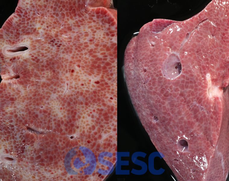
Detail of section of two livers: healthy (right) and affected (left) . In the healthy liver, the hepatic lobules were finely delimited by scant fibrous tissue. In contrast, the affected liver showed multifocal irregular areas of fibrous tissue deposition, which circumscribde and compressed the adjacent lobules (chronic interstitial hepatitis).
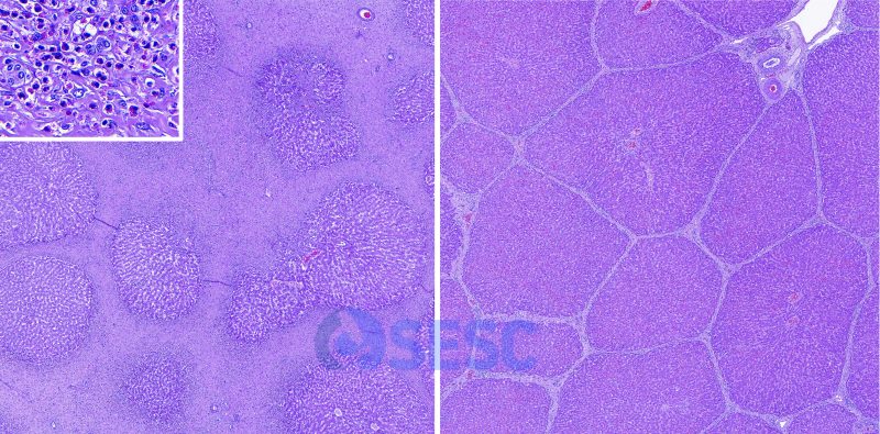
Histological section of the livers. On the right (normal liver), scant fibrous connective tissue can be observed between portal tracts. In contrast, milk spotted liver (on the left) presents multifocal areas of fibrosis that envelope the lobules, with abundant eosinophilic infiltration (inset).
