What lesions does the Rift Valley Fever virus cause?
Rift Valley fever is an emerging zoonotic viral disease (endemic to the African continent) transmitted by mosquito bites (mainly by species of the genera Aedes and Culex, both present in Catalonia) and through contact with tissues and fluids from infected animals.
Despite affecting different species, ruminants are particularly susceptible, especially sheep with high mortality levels. In affected animals, the clinical outcome is variable, nonspecific, and usually presents with fever and abortion in pregnant animals. In disease outbreaks, infected animals could reach the slaughterhouse, with viremia but subclinical, without any clinical and/or pathological alterations.
The most significant findings in animals suffering from the disease are miliary and generalised areas of necrosis (reddish and / or whitish grayish) in the liver (necrotizing hepatitis), as well as edema and hemorrhage of the gallbladder wall, hemorrhages in the mucosa of abomasum, splenomegaly, and generalized lymphadenomegaly with petechiae. Digested blood can also be seen in the gastrointestinal tract.
In case of suspicion, it is important to send fresh liver samples to the laboratory (for PCR) and also samples of tissues with visible lesions (histopathology) to confirm or rule out the suspicion. Although the probability of infection is low, it is important to use personal portective equipment such as gloves, mask and goggles when taking samples or handling these carcases. (AC)
The following images were obtained as part of an experimental infection carried out by the IRTA-CReSA high biocontainment unit.
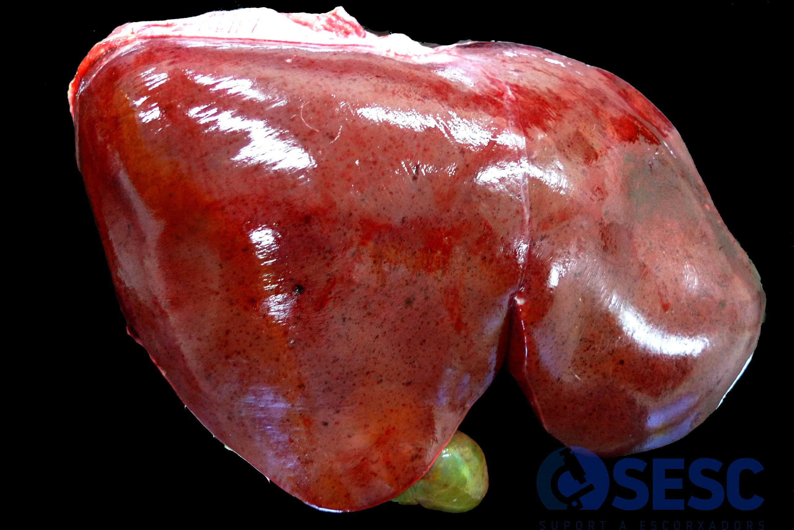
Liver. Ewe. Over the hepatic surface multifocal to generalized miliary areas showing darkish coloration can be seen, which correspond to foci of necrotizing hepatitis.
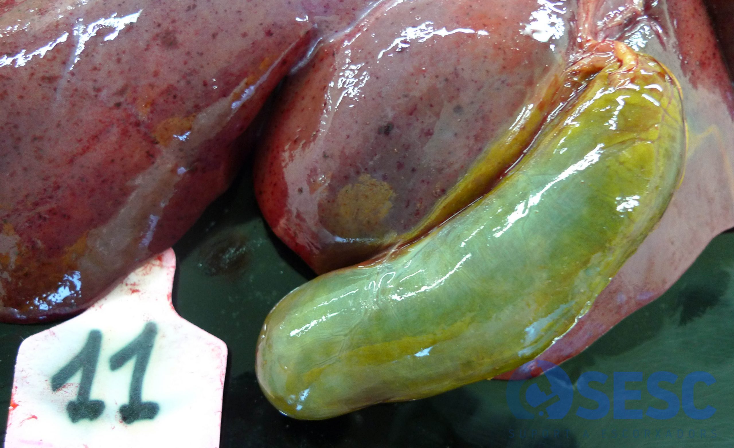
Liver. Ewe. The gallbladder is swollen and when sectioned (not shown in this image) its wall shows an increase in its thickness and a gelatinous appearance, which is consistent with gallbladder wall edema.
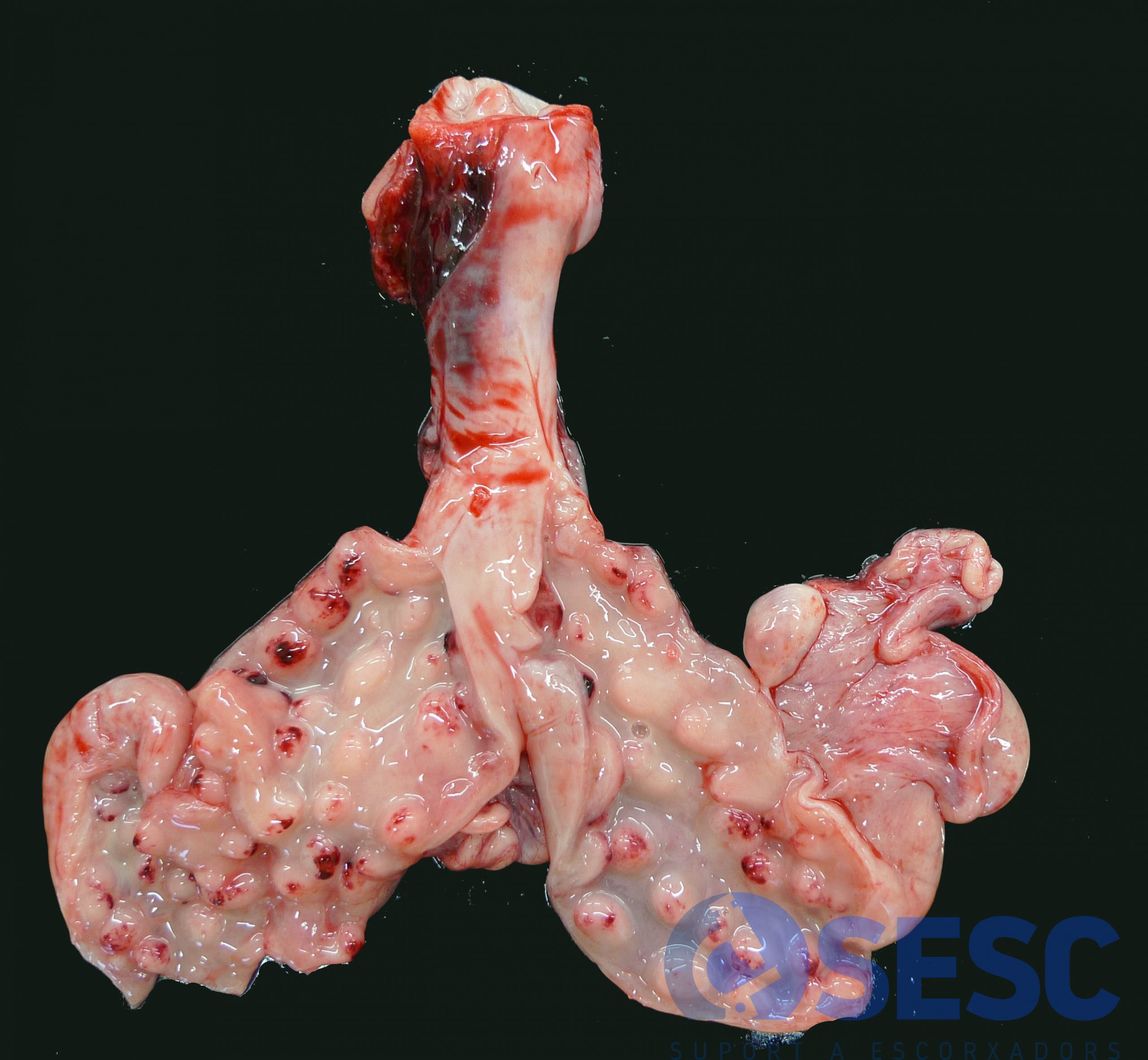
Uterus. Ewe. Multifocal hemorrhages in the endometrium, localized within the caruncles.
Heart. Ewe. This case presents endocardial equimoses.
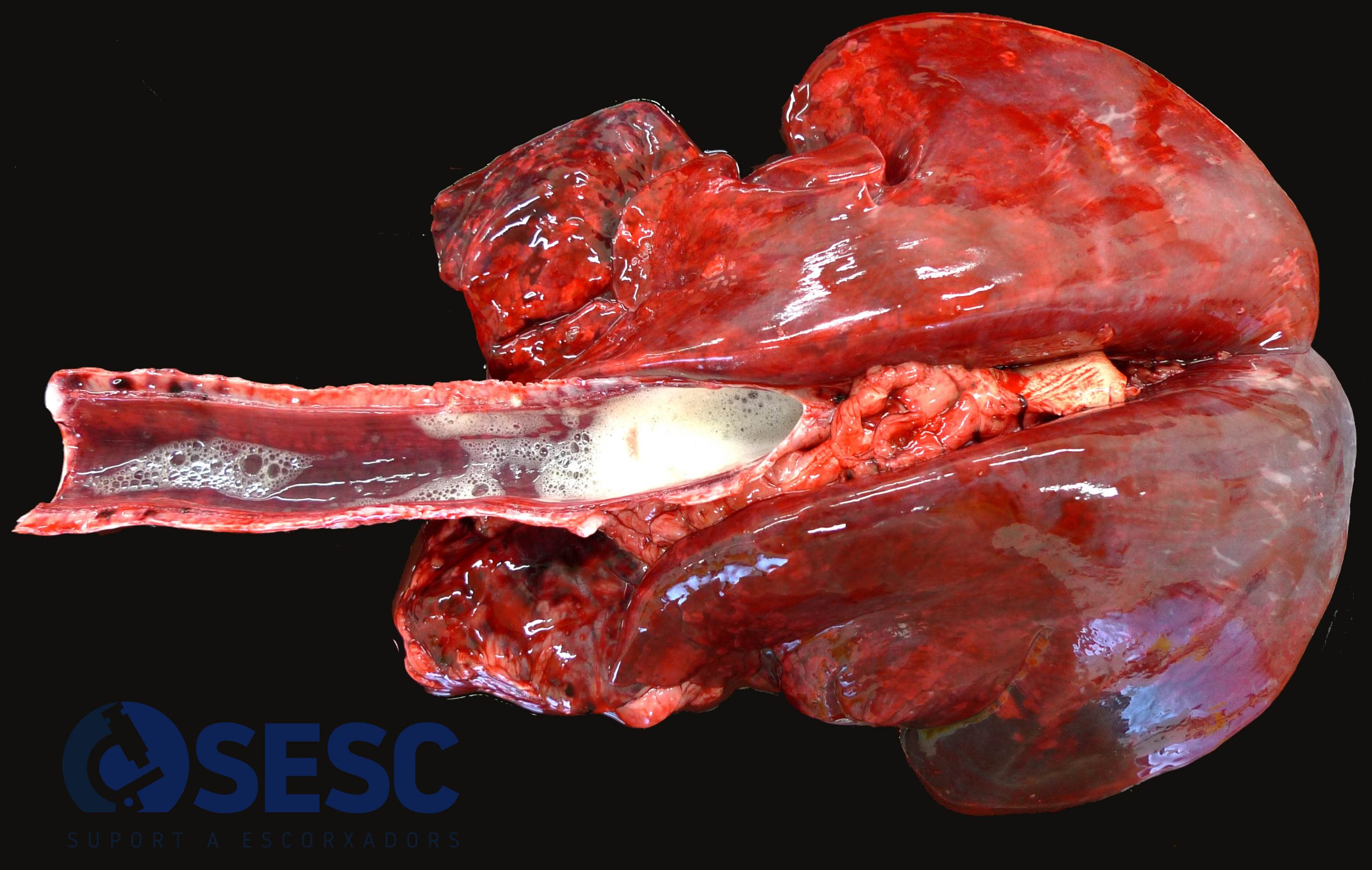
Lung. Ewe. The presence of foam in the tracheal lumen and the lack of pulmonary collapse are derived from a marked pulmonary edema and congestion. This case also shows pulmonary cranioventral consolidation.
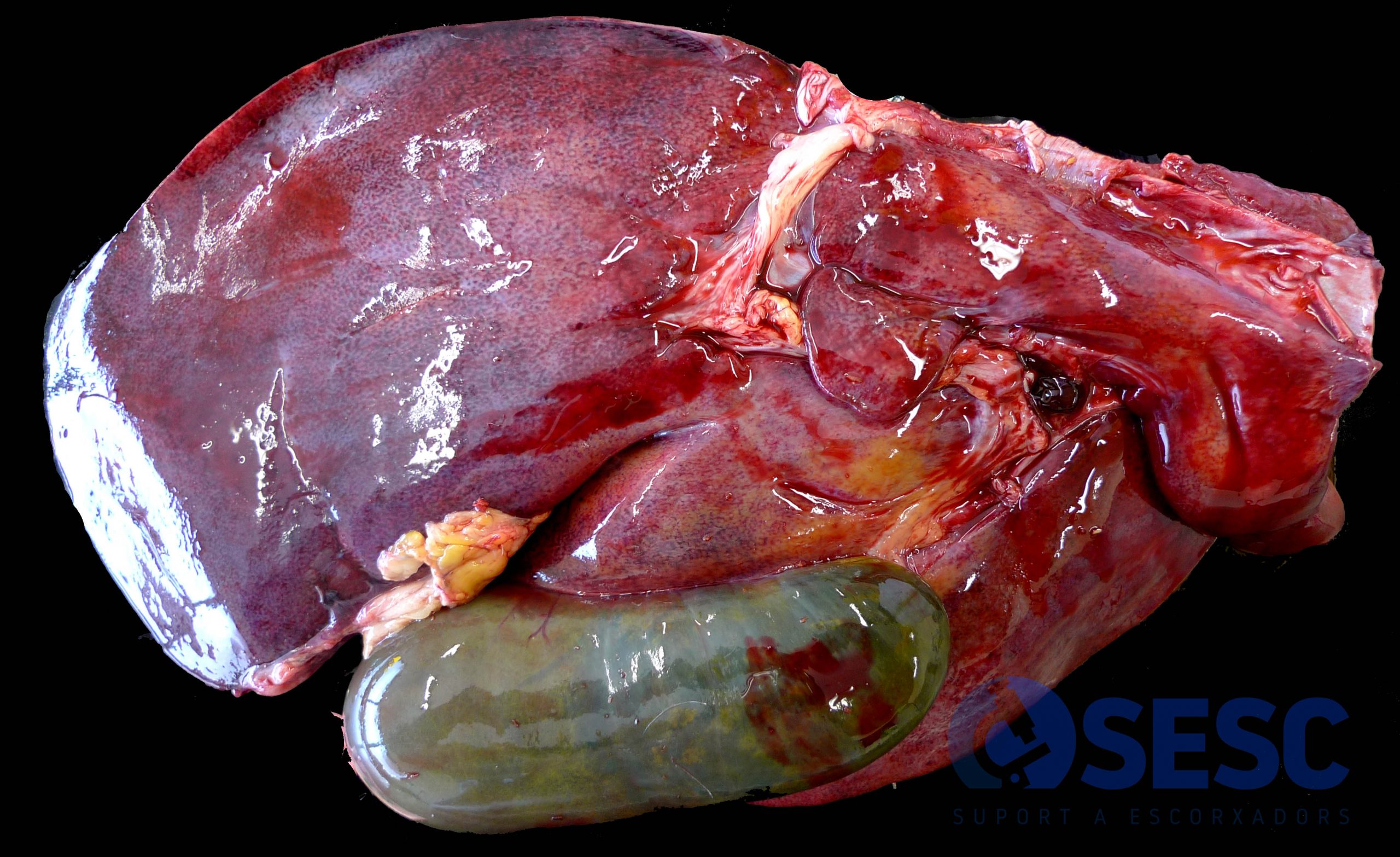
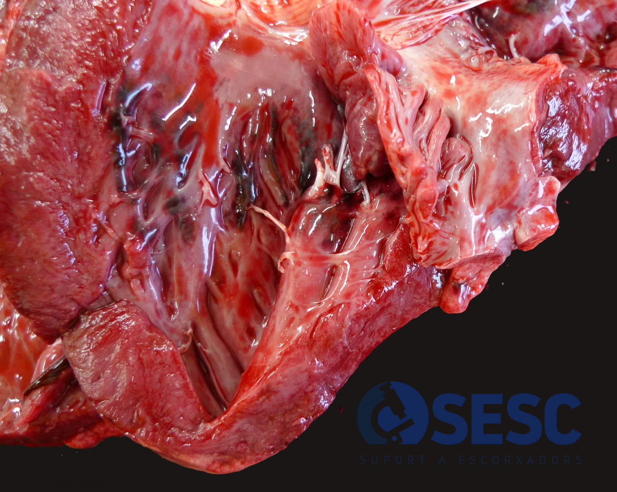
Liver. Ram. Hepatic lesion like the one shown above. This case shows a hemorrhage within the gallbladder wall.
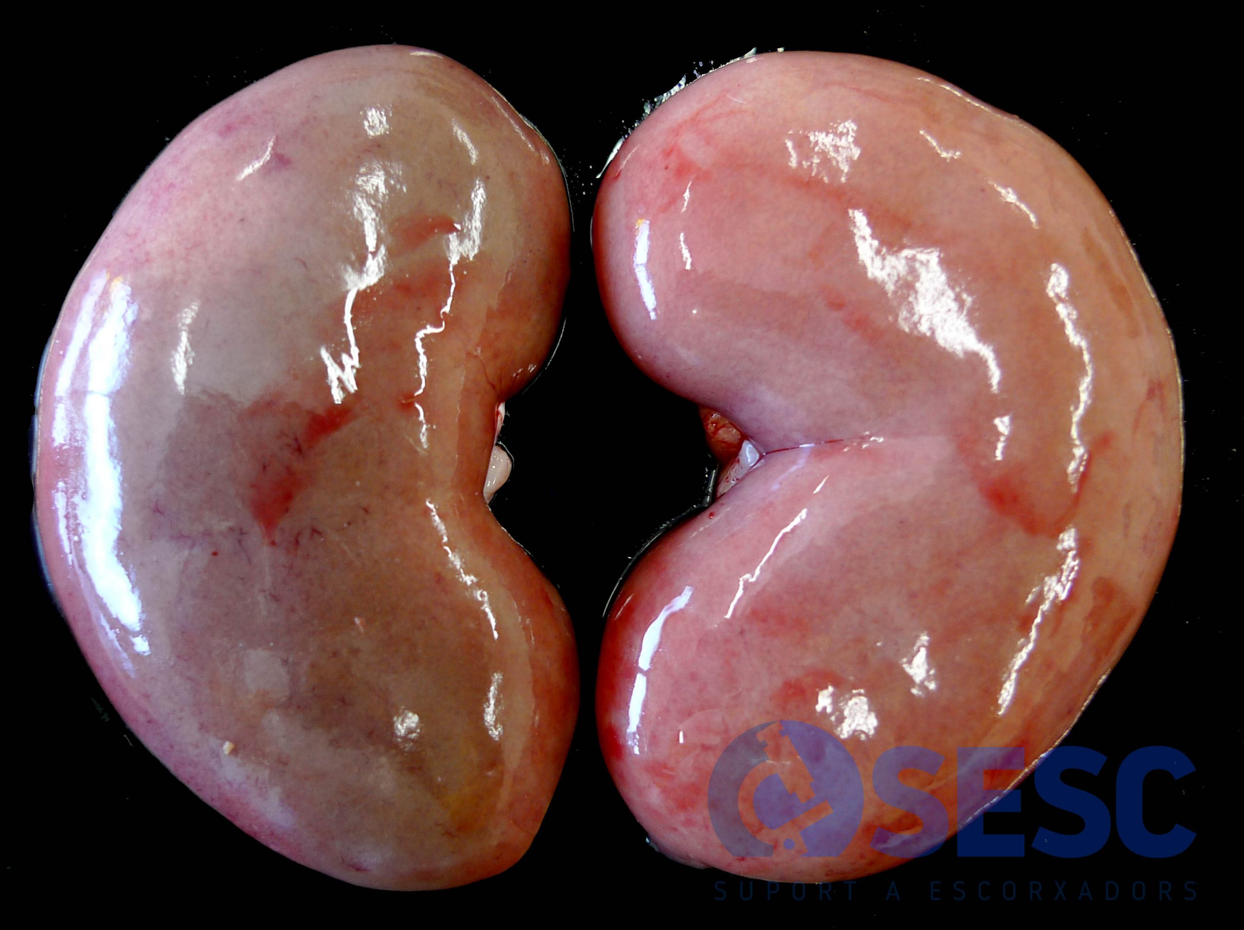
Kidney. Ram. Scant petechiae can be seen over the renal surface.
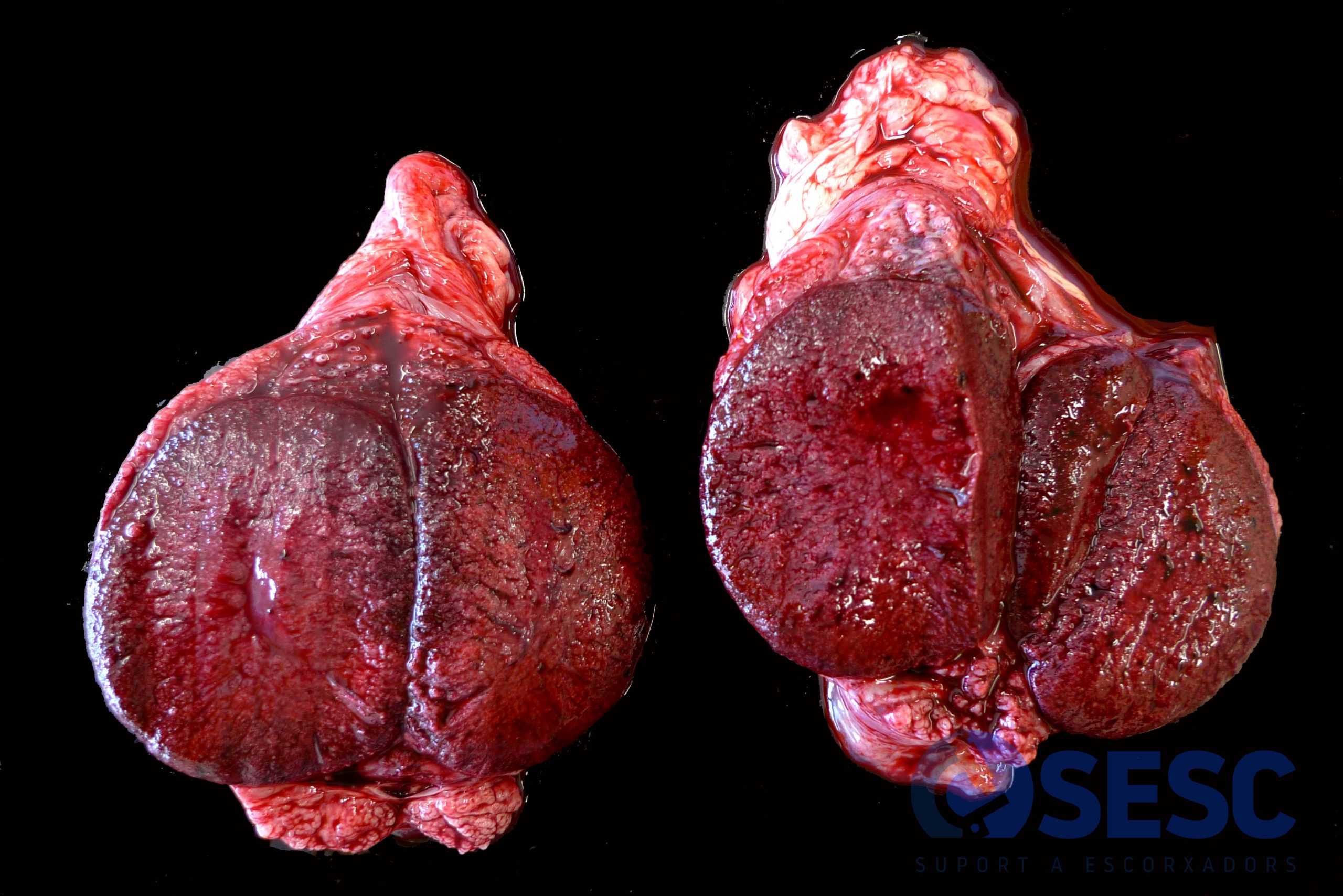
Testicles. Ram. Although it is not a commonly described lesion in the bibliography, this case presented intensely reddish coloration in both testicles (hemorrhagic).
