Bovine cysticercosis lesions in the heart, can the valves be involved?
One of the duties of official meat inspectors in ruminant slaughterhouses is bovine cysticercosis survilleance, which is a zoonotic disease. Cysticercus bovis is the intermediate form, and parasites striated muscle of bovines. Humans develop the adult parasite (Taenia saginata) in the gastrointestinal tract in case of ingesting meat with viable cysticercus. Surveillance of bovine cysticercosis in the slaughterhouse is thus a matter of public health.
In the muscles, the cysticerci appear as a cyst with a wall of variable thickness. The immune reaction of the host eliminates the parasite between 1 and 9 months following the infestation. After this, a focal granuloma with mineralized debris can be observed.
The cysticercus parasites the striated muscle of bovines and usually aims for the better oxygenated muscles (masticatory muscles, diaphragm, heart…) and these are the sites that should be inspected for lesions. The heart is a critical location, as it can show other macroscopical alterations that could rise the suspicion of a cysticercosis. One example is the myocardial adenomatoid tumour, of which we have had some cases that raised the suspicion of cysticercosis, but they are uncommon.
The presence of serous valvular cysts in the cardiac valves is more frequent (in a study with 30.000 bovines from slaughterhouse a prevalence of 11% was reported). These valvular cysts affect the conjunctive tissue of the valve, are incidental and it is believed they can derive from ecstatic lymph or blood vessels.
In contrast, cysticercosis exclusively affects muscular tissue. Occasionally it may be located in the subendocardial muscular tissue, hence appearing in the papillary musculature near to the valvular cords, but not in the valves. At SESC we have received some samples of valvular cysts, as well as cysticercosis near the valves, and we made a compilation of these cases in this post. (AC)
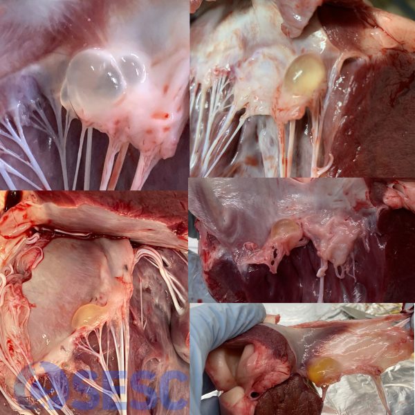
Examples of serous valvular cysts submitted to SESC. All of them affect the free leaflet of the cardiac valve. They can affect both the right and the left atrioventricular valves.
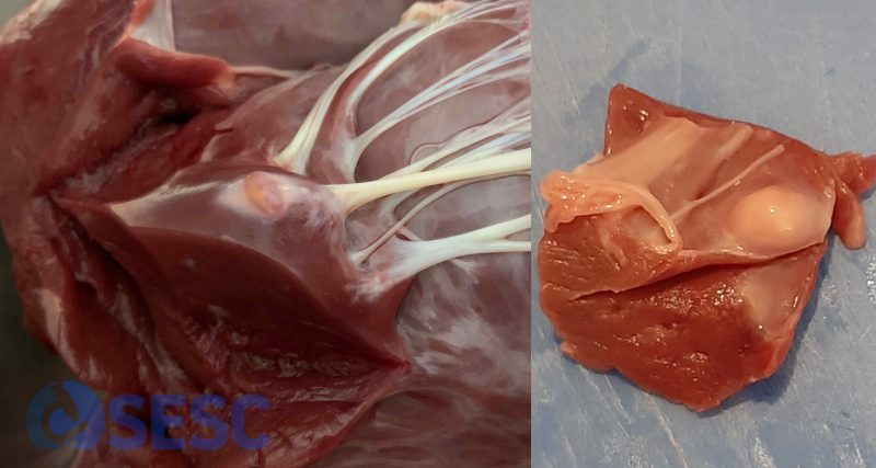
Examples of subendocardial cysticercosis submitted to SESC. Although they are located near the valvular cords, they always involve the cardiac muscle to some extent, hence they can’t be located in the free leaflet of the atrioventricular valves.
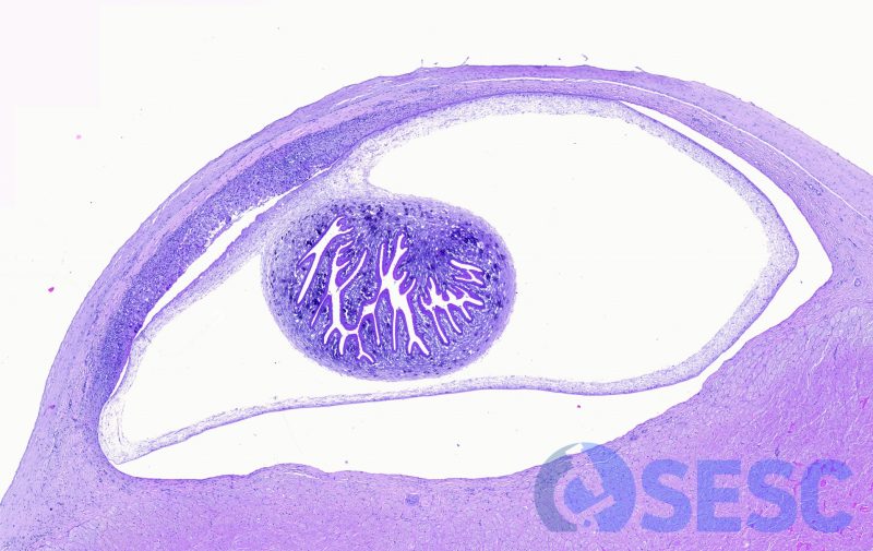
Histological section of the cysticercus shown previously (on the left in the previous image). Notice its subendocardial location, but in contact with the muscular tissue. The parasitic membrane can be observed, as well as the invaginated protoscolex.
